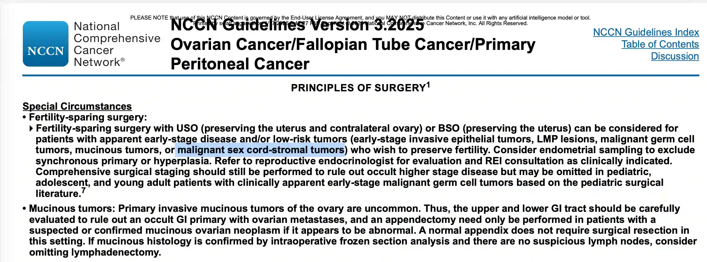
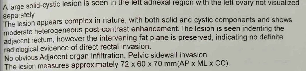
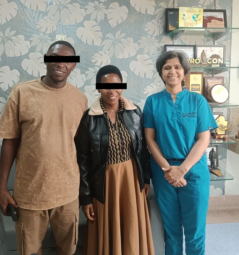
Preserving future fertility is an important consideration when treating ovarian tumours in children, adolescents and young women. However, fertility-preserving surgery (FSS) is appropriate only in carefully selected patients. The decision depends on the type of ovarian tumour, the extent of disease and accurate surgical staging.
⦿ In this 12-year-old girl treated at the Indian Ovarian Cancer Institute (IOCI), Ahmedabad, the ovarian tumour was removed intact and immediately examined by frozen section, a rapid pathological assessment performed while the patient remained under anaesthesia. Frozen section suggested a malignant sex cord stromal tumour, allowing the surgical team to confidently proceed with fertility-preserving surgery by preserving the uterus and the opposite ovary while completing comprehensive surgical staging, including peritoneal biopsies, omentectomy, pelvic and retroperitoneal lymph node dissection. Final histopathology confirmed a Juvenile Granulosa Cell Tumour confined to the left ovary (FIGO Stage IA) with negative peritoneal biopsies, omentum and 41 negative lymph nodes.
Fertility-preserving surgery is not suitable for every ovarian cancer. It is considered only when the tumour type and stage indicate that preserving the uterus and the opposite ovary is oncologically safe. Frozen section provides the intraoperative diagnosis that helps surgeons make this decision immediately, allowing both appropriate cancer surgery and preservation of future fertility whenever medically feasible.
⦿ Choosing the most appropriate surgical approach is an important step in ovarian cancer management. In this case, imaging suggested a localized left ovarian tumour without obvious distant spread. To assess the abdominal cavity directly, staging laparoscopy was performed before proceeding with definitive surgery.
⦿ During laparoscopy, surgeons identified an approximately 8 cm left ovarian mass with surface involvement, a small amount of pelvic fluid and tiny pelvic peritoneal deposits. Based on these findings, the surgical team converted to an open procedure to perform comprehensive staging surgery. Frozen section examination of the ovarian mass confirmed invasive malignancy during the operation, allowing complete staging to be performed in the same sitting.
Staging laparoscopy provides valuable real-time information about disease extent. In selected patients, it helps surgeons choose the safest and most appropriate operative strategy instead of relying only on imaging findings.
This clinical experience is shared by Indian Ovarian Cancer Institute (IOCI), Ahmedabad.
Every patient is unique. Treatment recommendations are individualized based on clinical evaluation, imaging, pathology and multidisciplinary discussion.

⦿ Not every patient with suspected advanced ovarian cancer should undergo immediate major surgery. In one of our recent cases at the Indian Ovarian Cancer Institute (IOCI), Ahmedabad, CT scan showed bilateral ovarian masses, ascites, omental involvement, and peritoneal deposits, suggesting advanced disease.
⦿ To better understand the disease and plan the safest treatment, our team first performed diagnostic laparoscopy. This allowed direct visualization of disease spread, PCI assessment, tissue biopsy, and individualized treatment planning before deciding the next steps.
⦿ Selecting the right treatment often begins with selecting the right investigation.
⦿ Not every patient with advanced ovarian cancer undergoes surgery immediately after diagnosis. In this case, the patient first received three cycles of neoadjuvant chemotherapy with Carboplatin and Paclitaxel before undergoing planned interval cytoreductive surgery. Follow-up imaging documented reduction in the size of both adnexal masses compared with the initial scans, after which surgery was scheduled.
⦿ During surgery, disease involving the ovaries, pelvic peritoneum, omentum and other abdominal surfaces was addressed through comprehensive cytoreduction followed by HIPEC. Histopathology later demonstrated residual viable high-grade serous carcinoma with a Chemotherapy Response Score (CRS2), indicating a moderate pathological response to preoperative chemotherapy.
⦿ Clinical Insight: In selected patients with advanced ovarian cancer, neoadjuvant chemotherapy can reduce tumour burden and may improve the feasibility of achieving optimal cytoreductive surgery. The final decision depends on imaging findings, overall fitness, disease distribution and multidisciplinary evaluation.
⦿ This clinical experience is shared by Indian Ovarian Cancer Institute (IOCI), Ahmedabad.
⦿ Every patient is unique. Treatment recommendations are individualized based on clinical evaluation, imaging, pathology and multidisciplinary discussion.

A recently managed case at Indian Ovarian Cancer Institute (IOCI) demonstrates what carefully selected minimally invasive surgery can achieve.
A 42-year-old woman with recurrent vaginal bleeding underwent total laparoscopic hysterectomy with right salpingo-oophorectomy and left salpingectomy. Laparoscopic assessment showed a bulky uterus but no free fluid, no enlarged pelvic lymph nodes and only small bilateral simple ovarian cysts.
⦿ Blood loss: only 10 mL
⦿ No blood transfusion
⦿ Ureter preserved during dissection
⦿ Specimen removed en masse
⦿ Secure laparoscopic vaginal cuff closure
The postoperative course was equally encouraging. The Foley catheter was removed on postoperative day 1, recovery was uneventful, and the patient was discharged ambulatory and haemodynamically stable.
Minimally invasive surgery is not simply about making smaller incisions.
When appropriately selected and performed by an experienced team, laparoscopy can combine precise pelvic dissection, very low blood loss, reduced surgical trauma and early postoperative recovery.
The goal is not only to treat the disease effectively—but also to help the patient return to normal life sooner.
Treatment planning after ovarian cancer surgery often depends on new imaging findings. In this case, PET-CT demonstrated two metabolically active lesions—one adjacent to the rectosigmoid region and another in the suprapubic region. The patient also had a recent diagnosis of endometrioid ovarian carcinoma following previous surgery, prompting multidisciplinary evaluation for possible residual disease.
Based on these findings, cytoreductive surgery was planned to evaluate and remove suspected disease. During surgery, suspicious areas involving the pelvic peritoneum, previous surgical scar, infundibulopelvic ligament region, vault and omentum were excised. Histopathology later showed that these suspicious areas represented chronic inflammation, foreign body giant cell reaction, suture granuloma and postoperative tissue changes, with no evidence of viable tumour cells.
Clinical Insight:
PET-CT is an important tool for identifying suspicious lesions, but imaging alone cannot confirm active cancer. Surgical assessment and histopathological examination remain the gold standard for distinguishing residual malignancy from postoperative inflammatory changes.
This clinical experience is shared by Indian Ovarian Cancer Institute (IOCI), Ahmedabad.
Every patient is unique. Treatment recommendations are individualized based on clinical evaluation, imaging, pathology and multidisciplinary discussion.

Swasth Parivar: twice a month, practical advice on recovery, diet and warning signs. No advertising. Stop any time.
Your number is used for these updates and for the clinic to reach you. It is never sold or passed on. To stop, reply STOP on WhatsApp.
Department
Shah’s Gastro, Cancer & Robotic Surgery Centre
SF-203, 2nd Floor
Olive Greens, Gota
S G Highway, Ahmedabad
Gujarat, India 382481
Google map link:
https://maps.app.goo.gl/2wawX5nnDgjkKoeZA?g_st=iw
Apollo Hospital I.L
Plot No. 1 A,
Apollo Hospital International Limited,
GIDC Bhat, Industrial Estate,
Gandhinagar, Gujarat 382428
Google map link:
https://maps.app.goo.gl/jRYUHgH6rFdGsk5g8