


Case Library - Robotic/ Laparoscopic Ovary surgery
Bilateral Ovarian Cysts Do Not Always Mean Ovarian Cancer
A recently managed case at Indian Ovarian Cancer Institute (IOCI) highlights the importance of careful evaluation before labelling an ovarian lesion as malignant.
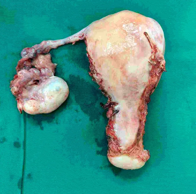
A 42-year-old woman presented with recurrent vaginal bleeding. MRI showed two well-defined simple cysts in both ovaries, with no solid enhancing nodule, ascites or enlarged pelvic lymph nodes. At laparoscopy, the ovarian cysts were small—approximately 3 × 2 cm on the right and 2 × 2 cm on the left—with no free fluid or enlarged pelvic nodes.
Surgery was tailored to the clinical situation: total laparoscopic hysterectomy with right salpingo-oophorectomy and left salpingectomy.
Final histopathology provided the definitive answer: the right ovarian lesion was a fibroma with a follicular cyst, with no malignant etiology.
The learning point
An ovarian cyst on imaging is not synonymous with ovarian cancer. The morphology of the lesion, presence or absence of solid components, ascites, peritoneal disease, lymphadenopathy and the overall clinical picture must all be considered before deciding the extent of surgery.
Previous Cancer Does Not Mean Every New Pelvic Lesion Is Recurrence
A recently managed case at Indian Ovarian Cancer Institute (IOCI) demonstrates an important principle in cancer care: a history of malignancy should raise suspicion—but should never replace tissue diagnosis.
The patient had a previous right renal PEComa (perivascular epithelioid cell tumour) treated with nephrectomy and was on systemic therapy. She subsequently presented with menorrhagia. Imaging demonstrated bilateral ovarian cysts, creating an important diagnostic question in a patient with a previous malignancy.
Instead of assuming that the ovarian findings represented metastatic or recurrent disease, the patient underwent definitive laparoscopic surgery.
- The operative findings were reassuring:
⦿ Bilateral small simple ovarian cysts
⦿ No free fluid
⦿ No enlarged pelvic lymph nodes
⦿ No obvious pelvic malignant deposits
- Histopathology subsequently showed:
⦿ Right ovarian fibroma
⦿ Follicular cyst
⦿ Adenomyosis
⦿ Leiomyoma
⦿ No malignant etiology
The learning point
In a patient with a previous cancer, every new lesion deserves evaluation—but not every lesion is recurrence.
Overdiagnosing recurrence can lead to unnecessary extensive treatment. Careful imaging, clinical assessment, appropriate surgery and histopathological confirmation help ensure that treatment is based on the actual disease, not simply the patient’s cancer history.
Anticoagulation Is an Important Part of Preoperative Cancer Planning
A recently managed case at Indian Ovarian Cancer Institute (IOCI) also highlights an often-overlooked aspect of cancer surgery: the patient’s medications and previous medical history can significantly influence surgical planning.
This patient had a history of right renal PEComa treated with nephrectomy and was receiving everolimus and apixaban. She presented with significant abnormal uterine bleeding/menorrhagia.
- Before undertaking surgery, the complete clinical picture had to be considered—including:
⦿ Previous major cancer surgery
⦿ Current systemic treatment
⦿ Anticoagulation with apixaban
⦿ Risk of perioperative bleeding
⦿ Need for definitive treatment of the uterine bleeding
The patient underwent laparoscopic surgery with minimal blood loss of only 10 mL and no blood transfusion. Postoperative recovery was uneventful, and she was ambulatory and haemodynamically stable at discharge.
The learning point
Safe cancer surgery begins before entering the operating room.
A detailed preoperative assessment—including medications, anticoagulants, previous treatments and medical comorbidities—is essential to balance bleeding risk, thrombotic risk and surgical benefit.
Good outcomes depend not only on the surgical technique, but also on careful multidisciplinary perioperative planning.
Minimally Invasive Surgery Can Mean Less Blood Loss and Faster Recovery
A recently managed case at Indian Ovarian Cancer Institute (IOCI) demonstrates what carefully selected minimally invasive surgery can achieve.
A 42-year-old woman with recurrent vaginal bleeding underwent total laparoscopic hysterectomy with right salpingo-oophorectomy and left salpingectomy. Laparoscopic assessment showed a bulky uterus but no free fluid, no enlarged pelvic lymph nodes and only small bilateral simple ovarian cysts.
- The operation was completed laparoscopically with:
⦿ Blood loss: only 10 mL
⦿ No blood transfusion
⦿ Ureter preserved during dissection
⦿ Specimen removed en masse
⦿ Secure laparoscopic vaginal cuff closure
The postoperative course was equally encouraging. The Foley catheter was removed on postoperative day 1, recovery was uneventful, and the patient was discharged ambulatory and haemodynamically stable.
The learning point
Minimally invasive surgery is not simply about making smaller incisions.
When appropriately selected and performed by an experienced team, laparoscopy can combine precise pelvic dissection, very low blood loss, reduced surgical trauma and early postoperative recovery.
The goal is not only to treat the disease effectively—but also to help the patient return to normal life sooner.

A Complex Ovarian Mass on Imaging: What Features Should Raise Suspicion?
A recently managed case at the Indian Ovarian Cancer Institute (IOCI) highlights an important principle: imaging can identify an ovarian mass as suspicious, but imaging alone cannot tell us exactly what the tumour is.
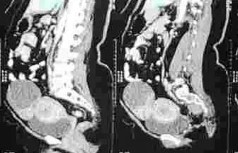
- What did the imaging show?
Ultrasound demonstrated a large complex cystic-solid lesion measuring approximately 8.5 × 11.3 cm in the right adnexa. The right ovary was not separately visualised, making a right ovarian origin likely. There was no ascites, significant lymphadenopathy or obvious distant disease on ultrasound.
- CECT abdomen and pelvis subsequently showed:
⦿ Large 11 × 9.2 cm solid-cystic mass anterior to the uterine fundus
⦿ Multiple internal septations
⦿ Enhancing solid component
⦿ Enhancing septae
⦿ Close proximity to the uterine fundus
⦿ Right ovary not separately identified
⦿ Possibility of right ovarian origin
⦿ No obvious liver, pancreatic or other visceral lesion
⦿ No significant retroperitoneal or mesenteric lymphadenopathy
⦿ No ascites/free fluid reported on the ultrasound evaluation
⦿ The imaging appearance therefore warranted evaluation for ovarian malignancy / malignant transformation.
What features of an ovarian cyst or mass are concerning?
When evaluating an adnexal lesion, imaging should systematically assess:
1. Cystic versus solid nature
A predominantly simple cyst is very different from a complex lesion containing substantial solid tissue.
2. Septations
Thin, smooth septations may be seen in benign lesions, whereas irregular or thickened septations increase concern.
3. Solid component
A true solid component, particularly when it enhances after contrast, is an important suspicious feature.
4. Papillary projections / mural nodules
These are important features in risk stratification. O-RADS defines a solid component as tissue protruding ≥3 mm into the cyst lumen; a papillary projection is a solid component surrounded by fluid on three sides. (American College of Radiology)
5. Internal vascularity
Doppler vascularity within solid tissue adds important information. O-RADS incorporates colour Doppler vascularity into adnexal lesion risk assessment. (American College of Radiology)
6. Irregular walls or septations
7. Ascites and peritoneal nodules
8. Bilaterality
9. Size and morphology
10. Evidence of disease outside the ovary
Such as omental/peritoneal deposits, enlarged nodes, pleural effusion or distant metastases.
Ultrasound is the primary imaging modality for characterization of most adnexal lesions, while MRI can provide additional soft-tissue characterization when ultrasound is indeterminate. (American College of Radiology)
But imaging cannot give the final tumour diagnosis.
In this patient, the tumour markers were not suggestive of epithelial ovarian cancer:
CA-125: 6.9 U/mL
CA 19-9: 0.6 U/mL
CEA: 2 ng/mL
Yet the final diagnosis was an adult-type granulosa cell tumour.
This is why:
A suspicious ovarian mass should not be labelled benign or malignant based on imaging or CA-125 alone.
When surgery is appropriate, careful surgical removal with intraoperative frozen section can establish the histologic direction and help determine the appropriate extent of surgery; the final diagnosis is confirmed on permanent histopathology.
Imaging tells us how suspicious the mass is.
Surgery and pathology tell us what it actually is.
Normal CA-125 Does Not Exclude Ovarian Cancer — Think About Inhibin
A recently managed case at the Indian Ovarian Cancer Institute (IOCI) demonstrates why ovarian tumour evaluation should not depend on CA-125 alone.
The patient had a large right ovarian solid-cystic mass with enhancing solid areas and septations. CA-125, CA 19-9 and CEA were not elevated.
When the morphology raises the possibility of a sex-cord stromal tumour, the tumour marker panel needs to be expanded.
In this case, Inhibin B was specifically tested and was >964.0, providing an important clue toward granulosa cell tumour.
- The final histopathology confirmed:
Adult-type granulosa cell tumour of the right ovary.
The tumour measured 12.5 × 11.5 × 6 cm, with the ovarian capsule intact. There was no ovarian surface involvement, no omental or peritoneal tumour, and all 29/29 regional lymph nodes were negative. Final pathological staging was pT1a pN0.
- Why is Inhibin important?
Granulosa cell tumours are sex-cord stromal tumours, biologically different from the much more common epithelial ovarian cancers.
Therefore, depending on the imaging and clinical suspicion, evaluation may include markers such as:
• Inhibin A / Inhibin B
• AMH in appropriate clinical settings
• CA-125 and other epithelial markers when clinically indicated
The key lesson:
Tumour markers are not interchangeable.
A normal CA-125 does not mean that an ovarian mass is benign.
If imaging shows a large complex ovarian mass with solid tissue, septations or enhancement, the tumour type must be considered before deciding which markers are useful.
Correct diagnosis begins with asking the correct question:
“What type of ovarian tumour could this be?”
Why an Ovarian Tumour Must Be Removed Intact
A recently managed case at the Indian Ovarian Cancer Institute (IOCI) demonstrates one of the most important principles in ovarian cancer surgery:
Avoid rupture of a suspicious ovarian tumour.
The patient had a large right adnexal mass. At surgery, the mass was approximately 10 × 8 cm. It was carefully dissected circumferentially and removed en masse, without compromising its capsule.
Frozen section suggested a granulosa cell tumour, allowing the surgical team to proceed with appropriate oncological staging.
- Final pathology showed:
• Adult-type granulosa cell tumour
• Tumour size 12.5 × 11.5 × 6 cm
• Capsule intact
• No ovarian surface involvement
• No fallopian tube surface involvement
• Omentum negative
• Peritoneal biopsy negative
• Contralateral ovary negative
• 0/29 lymph nodes involved
• Final stage pT1a pN0
Why is avoiding rupture so important?
For an apparently confined ovarian tumour, rupture can result in tumour cells being released into the peritoneal cavity and may alter the pathological stage.
Therefore, the surgical objective is not merely:
“Remove the ovarian cyst.”
It is:
“Remove the ovarian tumour intact, without spillage, while performing appropriate surgical staging.”
This is particularly important when imaging shows a large, complex, solid-cystic or potentially malignant ovarian mass.
In this case, intact removal, frozen section and complete staging established that the disease was confined to the ovary.
The message for patients:
A large ovarian cyst should not automatically be treated like a routine benign cyst.
If malignancy is possible, surgical technique matters.
Resection without rupture → frozen section → appropriate staging → final histopathology
That sequence can make a major difference in ovarian cancer management.
Robotic Ovarian Cancer Surgery: Minimally Invasive Does Not Mean Less Cancer Surgery
A recently managed case at the Indian Ovarian Cancer Institute (IOCI) demonstrates how robotic surgery can be used not merely to remove an ovarian mass, but to perform comprehensive ovarian cancer staging.
The patient had a large suspicious right ovarian mass measuring approximately 10 × 8 cm at surgery. There was also a bulky uterus with an approximately 5 × 5 cm intramural fundal fibroid and a few enlarged bilateral pelvic lymph nodes. Importantly, there was no visible disease elsewhere in the abdomen and no free fluid.
The procedure was performed using the Da Vinci XI robotic surgical system.
- The surgery included:
• Robotic right adnexal mass excision
• Hysterectomy
• Left salpingo-oophorectomy
• Right salpingectomy
• Pelvic sentinel lymph-node identification using ICG
• Complete bilateral pelvic lymph-node dissection
• Retroperitoneal lymph-node sampling
• Infracolic omentectomy
• Peritoneal assessment/biopsy
• Frozen section of the ovarian mass
The final pathology confirmed adult-type granulosa cell tumour.
- Importantly:
29/29 lymph nodes were negative, the omentum was free of tumour, the peritoneal biopsy was negative and the final stage was pT1a pN0.
What does this case teach us about robotic ovarian cancer surgery?
Robotic surgery is not simply about:
“small cuts” or “faster recovery.”
In a properly selected patient, an experienced robotic pelvic oncologist can use the platform for:
precise pelvic dissection + lymph-node assessment + retroperitoneal dissection + omentectomy + complete surgical staging.
- The robotic platform can provide:
• Magnified three-dimensional vision
• Stable camera control
• Wristed instruments
• Precise dissection in confined pelvic spaces
• Improved ergonomics for complex pelvic surgery
The oncological completeness of the operation comes first.
The technology is valuable only when it allows the surgeon to perform the right cancer operation safely and completely.
Robotic surgery should not mean “less surgery.”
It should mean the appropriate cancer surgery through a minimally invasive approach, when the patient and disease are suitable.
Laparoscopic BSO: Definitive Ovarian Suppression With Early Recovery
⦿ A recently managed patient at Indian Ovarian Cancer Institute (IOCI), Ahmedabad underwent laparoscopic bilateral salpingo-oophorectomy (BSO) as part of management of hormone-receptor-positive metastatic breast cancer.
⦿ The procedure was performed through four laparoscopic ports. Both infundibulopelvic pedicles were carefully dissected, secured and divided, and the adnexa were removed in an endobag. Operative time was approximately 1 hour, with only 30 mL blood loss and no requirement for transfusion.
⦿ The postoperative recovery was also uncomplicated. The patient was ambulatory, comfortable, tolerating diet and haemodynamically stable, with a clean surgical wound at discharge.
⦿ This illustrates an important option for patients who require definitive ovarian function suppression.
⦿ Key learning: When surgical ovarian suppression is appropriate, a laparoscopic BSO can provide definitive treatment through a minimally invasive approach, with low blood loss, limited surgical trauma and early postoperative recovery.
⦿ For a patient requiring long-term ovarian suppression, this can also avoid the need for repeated visits for injectable therapy.
Why Remove Both Ovaries in Hormone-Receptor Positive Metastatic Breast Cancer?
⦿ A recently managed patient at Indian Ovarian Cancer Institute (IOCI), Ahmedabad with metastatic, hormone-receptor-positive breast carcinoma underwent laparoscopic bilateral salpingo-oophorectomy as part of her systemic cancer management.
⦿ In premenopausal women with hormone-sensitive breast cancer, ovarian estrogen production can contribute to tumour stimulation. Ovarian function suppression therefore forms an important component of endocrine treatment in appropriately selected patients.
⦿ Ovarian suppression can be achieved medically with GnRH agonist injections or surgically by removing the ovaries.
⦿ Surgical ovarian ablation provides definitive and irreversible ovarian suppression, whereas injectable treatment is reversible but requires ongoing administration.
Key learning:
BSO can be an important therapeutic option in selected hormone-receptor-positive breast cancer patients who require ovarian suppression. The decision should consider the patient’s age, systemic treatment plan, fertility wishes, preference and acceptance of permanent menopause.
Injectable Ovarian Suppression vs Laparoscopic BSO: A Practical Treatment Choice
For a premenopausal patient requiring ovarian function suppression, GnRH agonist injections and bilateral salpingo-oophorectomy (BSO) achieve the same broad therapeutic objective—reducing ovarian hormone production—but in very different ways.
- Injectable ovarian suppression
⦿ Reversible
⦿ Requires repeated injections
⦿ Requires continued adherence
⦿ Ovarian function may recover after treatment is stopped
- Laparoscopic BSO
⦿ Definitive and irreversible
⦿ One-time procedure
⦿ No repeated ovarian-suppression injections
⦿ Causes permanent loss of ovarian function
In this recently managed case at Indian Ovarian Cancer Institute (IOCI), Ahmedabad, BSO was performed laparoscopically through four ports. The procedure took approximately 1 hour, blood loss was 30 mL, and the patient had an uncomplicated early recovery.
Therefore, in a patient who has completed childbearing and requires long-term, reliable ovarian suppression, laparoscopic BSO may be a practical alternative to continuing injectable therapy.
Key learning: Neither approach is universally better. The choice should be individualized after discussing reversibility, permanent menopause, fertility, treatment duration, adherence and patient preference.
Grossly Normal Ovaries: Why Histopathology Still Matters
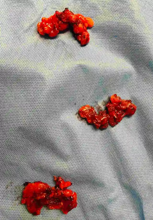
⦿ Intraoperatively, the uterus and both ovaries appeared grossly normal. Nevertheless, both adnexa were removed and submitted for histopathological examination. The specimens were entirely submitted for evaluation.
⦿ The final pathology showed a right ovarian follicular cyst, while the left ovary showed no significant pathology. Both fallopian tubes were unremarkable, and no malignancy was identified.
⦿ This demonstrates why the surgeon’s visual assessment cannot replace pathology.
⦿ A grossly normal ovary may contain an incidental benign lesion, while definitive assessment of removed tissue requires microscopic examination.
Key learning: In oncology surgery, proper specimen handling and complete histopathological examination are essential. Operative appearance describes what the surgeon sees; histopathology establishes what the tissue actually contains.
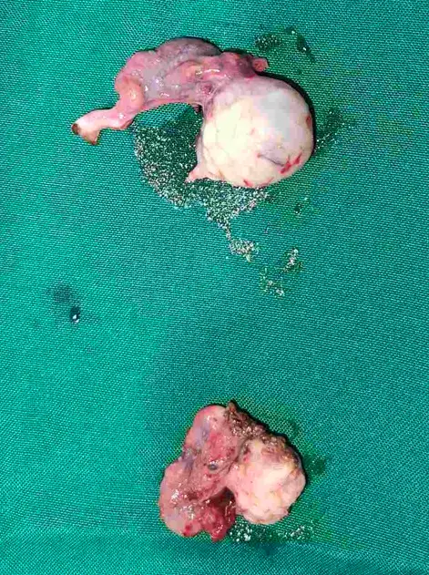
ROBOTIC COMPLETE STAGING FOR GRANULOSA CELL TUMOUR
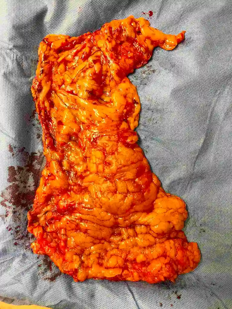
Key learning:
Robotic surgery can extend well beyond a simple hysterectomy or adnexal procedure. In appropriately selected ovarian malignancies, complete surgical staging can be performed robotically, combining pelvic surgery, lymphadenectomy, retroperitoneal dissection, omentectomy and peritoneal assessment in one procedure.
ROBOTIC RETROPERITONEAL LYMPH-NODE DISSECTION
Key learning:
Robotic retroperitoneal lymph-node dissection can make systematic ovarian cancer staging feasible through a minimally invasive approach when performed by an experienced pelvic oncology team.
ROBOTIC SURGERY: PRECISION WHEN MULTIPLE PROCEDURES ARE NEEDED
Key learning:
Robotic surgery combines precision, magnified visualization, wristed instrumentation and minimally invasive access, allowing complex staging procedures to be performed without automatically resorting to a large abdominal incision.
ROBOTIC OVARIAN CANCER SURGERY: HISTOPATHOLOGY COMPLETES THE STORY
Key learning:
Robotic technology + sound oncological principles + systematic histopathological evaluation together determine the quality of minimally invasive ovarian cancer surgery.
Get health updates from Dr Swati Shah on WhatsApp
Swasth Parivar: twice a month, practical advice on recovery, diet and warning signs. No advertising. Stop any time.
Your number is used for these updates and for the clinic to reach you. It is never sold or passed on. To stop, reply STOP on WhatsApp.