

Case Library - 3rd stage Ovary cancer
Advanced Ovarian Cancer with Ascites – Individualized Treatment Planning
⦿ Persistent abdominal swelling after menopause should never be ignored. At the Indian Ovarian Cancer Institute (IOCI), Ahmedabad, we recently evaluated a woman with progressive abdominal distension. Investigations revealed bilateral ovarian masses, markedly elevated CA-125, ascites, and CT findings suggestive of advanced ovarian cancer with peritoneal involvement.
⦿ Instead of proceeding directly to major surgery, our team performed diagnostic laparoscopy to accurately assess disease extent. The procedure included Peritoneal Cancer Index (PCI) assessment, peritoneal biopsy, and Pressurized Intraperitoneal Aerosol Chemotherapy (PIPAC) as part of individualized treatment planning.
⦿ Every patient with advanced ovarian cancer presents differently. Careful evaluation helps determine the most appropriate treatment strategy.
Indian Ovarian Cancer Institute (IOCI), Ahmedabad
Dedicated to ovarian cysts, complex ovarian tumours, ovarian cancer, CRS, HIPEC, PIPAC and robotic gynecologic oncology surgery.
Why Diagnostic Laparoscopy Was Chosen Before Major Ovarian Cancer Surgery
⦿ Not every patient with suspected advanced ovarian cancer should undergo immediate major surgery. In one of our recent cases at the Indian Ovarian Cancer Institute (IOCI), Ahmedabad, CT scan showed bilateral ovarian masses, ascites, omental involvement, and peritoneal deposits, suggesting advanced disease.
⦿ To better understand the disease and plan the safest treatment, our team first performed diagnostic laparoscopy. This allowed direct visualization of disease spread, PCI assessment, tissue biopsy, and individualized treatment planning before deciding the next steps.
⦿ Selecting the right treatment often begins with selecting the right investigation.
PIPAC for Peritoneal Surface Disease – A Specialized Treatment Approach
⦿ At the Indian Ovarian Cancer Institute (IOCI), Ahmedabad, selected patients with suspected advanced ovarian cancer and peritoneal disease may benefit from specialized evaluation using diagnostic laparoscopy.
⦿ In a recent case, laparoscopic assessment demonstrated widespread peritoneal involvement. During the same procedure, Pressurized Intraperitoneal Aerosol Chemotherapy (PIPAC) was administered after careful assessment and biopsy as part of the planned management strategy.
⦿ PIPAC is a specialized treatment option that may be considered for selected patients after multidisciplinary evaluation. Individualized treatment planning remains central to achieving the best possible outcomes.
High CA-125, Bilateral Ovarian Masses and Omental Caking on CT Scan – What Do These Findings Mean?
⦿ Many patients become anxious after reading CT scan reports mentioning bilateral ovarian masses, omental caking, peritoneal deposits, or after seeing a high CA-125 result. These findings can raise suspicion for advanced ovarian cancer, but treatment decisions should never be based on a single report alone.
⦿ At the Indian Ovarian Cancer Institute (IOCI), Ahmedabad, we evaluate imaging findings together with clinical examination, tumour markers, laparoscopic assessment and pathology before recommending treatment. In a recent patient with these findings, diagnostic laparoscopy and peritoneal biopsy helped guide further management.
Stage IIIC Ovarian Cancer with Gross Ascites & Poor Performance Status: Why Initial Evaluation by a Gynecologic Oncologist Matters
A recently managed case at the Indian Ovarian Cancer Institute (IOCI), Ahmedabad involved a 73-year-old woman with high-grade serous carcinoma of ovarian/Müllerian origin, gross ascites, omental infiltration, peritoneal deposits, supra-diaphragmatic nodes, paracardiac nodes, gastrohepatic deposits and poor performance status. After detailed evaluation by a Gynecologic Oncologist, Neoadjuvant Chemotherapy (9 weekly Paclitaxel + Carboplatin) was advised before Cytoreductive Surgery.
- Clinical Insight
Not every patient with Stage III ovarian cancer, peritoneal carcinomatosis or gross ascites should undergo immediate surgery.
- The first decision is often the most important:
⦿ Primary Cytoreductive Surgery
⦿ Neoadjuvant Chemotherapy (NACT) followed by Interval Cytoreductive Surgery
- This decision depends on:
⦿ Extent of disease
⦿ Performance status
⦿ Nutritional status
⦿ Medical co-morbidities
⦿ Anaesthetic fitness
⦿ Likelihood of achieving complete cytoreduction
Take-home message:
Patients with advanced ovarian cancer, peritoneal carcinomatosis, omental deposits or gross ascites should ideally be evaluated by a Gynecologic Oncologist / Ovarian Cancer Surgeon / Peritoneal Surface Malignancy Surgeon, as selecting the correct sequence of treatment significantly influences outcomes.
Preparing for Ovarian Cancer Surgery: Nutrition and Medical Optimization Are as Important as the Operation
A recently operated case at the Indian Ovarian Cancer Institute (IOCI), Ahmedabad was a 73-year-old patient with advanced ovarian cancer who received 9 cycles of Neoadjuvant Chemotherapy before surgery. Along with tumour response assessment, careful pre-operative optimisation was performed because of age, co-morbidities and respiratory illness before undertaking major Cytoreductive Surgery.
- Clinical Insight
Successful Cytoreductive Surgery begins long before entering the operation theatre.
- Many patients with advanced ovarian cancer develop:
⦿ Weight loss
⦿ Poor nutrition
⦿ Low protein levels
⦿ Reduced muscle mass
⦿ Weakness
⦿ Respiratory problems
⦿ Diabetes or hypertension
- Management often requires a multidisciplinary team including:
⦿ Gynecologic Oncologist
⦿ Intensivist
⦿ Clinical Nutrition Team
⦿ Chest Physician / Pulmonologist
⦿ Anaesthesiologist
⦿ Physician
Optimising nutrition and improving medical fitness before surgery reduces complications and helps recovery.
Take-home message:
Preparing the patient is as important as preparing the operation. A well-planned multidisciplinary approach can safely convert high-risk patients into suitable candidates for major ovarian cancer surgery.
Stage III Ovarian Cancer Is Primarily a Peritoneal Surface Disease — Not Always an Organ Removal Surgery
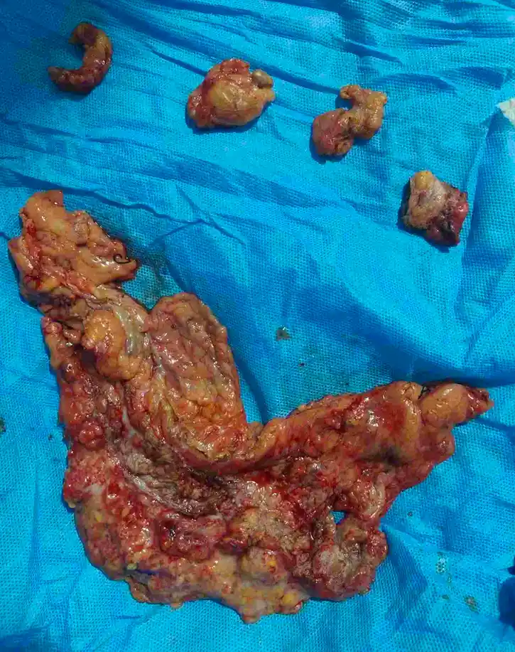
A recently operated case at the Indian Ovarian Cancer Institute (IOCI), Ahmedabad underwent Cytoreductive Surgery (CC1) after Neoadjuvant Chemotherapy. Operative findings included multiple peritoneal deposits, omental caking, pelvic peritoneal disease, cul-de-sac deposits, small bowel surface deposits, mesenteric deposits, falciform ligament deposits, paracolic peritoneal deposits and lesser omental deposits. Complete cytoreduction was achieved without major liver, stomach, spleen or pancreatic resection.
- Clinical Insight
Stage III ovarian cancer usually spreads over the surface of the abdomen (peritoneum) rather than deeply invading solid organs.
- Common operative findings include:
⦿ Peritoneal deposits
⦿ Omental caking
⦿ Pelvic peritoneal disease
⦿ Small bowel serosal deposits
⦿ Mesenteric deposits
⦿ Falciform ligament deposits
⦿ Paracolic peritoneal deposits
These deposits can often be removed by specialised peritonectomy procedures while preserving major abdominal organs whenever oncologically appropriate.
In elderly patients with multiple co-morbidities, careful anaesthesia planning, peri-operative monitoring and management at a high-volume ovarian cancer centre are equally important components of treatment.
Take-home message:
Advanced Stage III ovarian cancer frequently represents peritoneal surface disease. Successful Cytoreductive Surgery depends on specialised peritoneal surgery, experienced anaesthesia and multidisciplinary peri-operative care.
Recovery After Extensive Cytoreductive Surgery at 73 Years: The Importance of Team-Based Critical Care
A recently operated case at the Indian Ovarian Cancer Institute (IOCI), Ahmedabad involved a 73-year-old patient who underwent extensive Cytoreductive Surgery (CC1) for advanced ovarian cancer after Neoadjuvant Chemotherapy. Following surgery, intensive multidisciplinary care was required before the patient recovered, became ambulatory and was discharged in a stable condition.
- Clinical Insight
Recovery after major ovarian cancer surgery depends on much more than the operation itself.
- Some patients may require:
⦿ ICU care
⦿ Non-invasive ventilatory support (BiPAP), when indicated
⦿ Chest Physician support
⦿ Intensivist-led critical care
⦿ Cardiology evaluation
⦿ Respiratory physiotherapy
⦿ Nutrition support
⦿ Early mobilisation
⦿ Protocol-based postoperative management
Even elderly patients with significant co-morbidities can recover well when managed through coordinated multidisciplinary care.
Take-home message:
Advanced ovarian cancer treatment is a team effort. Experienced surgeons, anaesthesiologists, intensivists, chest physicians, cardiologists, nutrition experts and nursing teams together help patients recover safely and return to routine life after major Cytoreductive Surgery.
Diagnostic Laparoscopy in Advanced Ovarian Cancer: Surgery First or Chemotherapy First?
⦿ A recently managed case at the Indian Ovarian Cancer Institute (IOCI), Ahmedabad highlights an important step in advanced ovarian cancer treatment.
⦿ The patient had Stage IIIC high-grade serous ovarian cancer with moderate to gross ascites, bilateral ovarian disease, peritoneal and omental metastases, and a very high CA-125 (9709 U/mL).
⦿ Instead of directly proceeding to major surgery, Diagnostic Laparoscopy was performed.
⦿ During laparoscopy, the disease was assessed to determine whether Complete Cytoreductive Surgery (CC0/CC1) was achievable.
⦿ The most important finding was extensive disease over the small bowel serosa and mesentery, making complete tumour clearance unlikely at that stage.
⦿ Rather than performing an incomplete surgery, the patient was started on Neoadjuvant Chemotherapy (NACT) first.
- Why is Diagnostic Laparoscopy Important?
- It helps answer one crucial question:
Can all visible disease be removed safely?
If complete cytoreduction is unlikely,
✔ Neoadjuvant Chemotherapy is preferred.
If complete clearance appears achievable,
✔ Primary Cytoreductive Surgery is planned.
This approach avoids unnecessary major surgery while improving the chances of achieving complete tumour removal later.
Take-home message:
Diagnostic Laparoscopy is one of the most valuable decision-making tools in Stage III and Stage IV ovarian cancer, helping determine whether Primary Cytoreductive Surgery or Neoadjuvant Chemotherapy is the better first treatment.
Why Surgery is Still Needed After Excellent Response to Neoadjuvant Chemotherapy
A recently managed case at the Indian Ovarian Cancer Institute (IOCI), Ahmedabad demonstrated an excellent response to Neoadjuvant Chemotherapy.
- Initially, the patient had bilateral ovarian masses, moderate to gross ascites, omental deposits and peritoneal metastases . After chemotherapy:
✔ CA-125 reduced dramatically
✔ Ascites became minimal
✔ Ovarian lesions became much smaller
✔ CT showed only residual adnexal lesions with mild ascites
✔ During surgery, most deposits appeared as “burnt out deposits”, with only minimal free fluid remaining.
- Many patients ask:
“If the CT scan looks much better after chemotherapy, why is surgery still necessary?”
Because chemotherapy often leaves behind microscopic residual viable tumour cells.
These remaining cells can regrow after chemotherapy is completed and may eventually lead to recurrence.
Therefore, even after an excellent radiological response, Interval Cytoreductive Surgery (CRS) remains the standard treatment whenever complete tumour removal is feasible.
The goal is not only to shrink the disease—but to remove every remaining visible tumour.
Take-home message:
A good CT scan after chemotherapy is encouraging—but it does not mean all cancer has disappeared. Surgery remains an essential part of treatment to maximise long-term disease control.
Why the Pouch of Douglas (Cul-de-sac) is One of the Most Important Areas During Cytoreductive Surgery
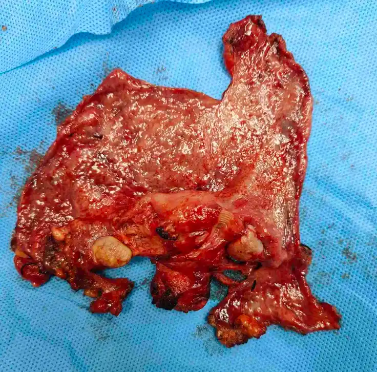
A recently managed case at the Indian Ovarian Cancer Institute (IOCI), Ahmedabad demonstrated disease involving the cul-de-sac (Pouch of Douglas) along with deposits over the rectal and sigmoid serosa, despite good response to chemotherapy. Complete cul-de-sac resection with pelvic peritonectomy was performed as part of Cytoreductive Surgery.
- Why is the Pouch of Douglas Important?
The Pouch of Douglas (Rectouterine pouch/Cul-de-sac) is the most dependent part of the pelvis.
Cancer cells floating within peritoneal fluid or ascites naturally settle here due to gravity.
Even when imaging shows a good response after chemotherapy, this area frequently harbours residual disease.
For this reason, experienced ovarian cancer surgeons carefully inspect and often remove the cul-de-sac peritoneum whenever involved, aiming for complete cytoreduction.
Missing disease in this region can leave behind residual tumour and reduce the effectiveness of surgery.
Take-home message:
Complete Cytoreductive Surgery is not simply removal of the ovaries. Careful assessment and clearance of the Pouch of Douglas, pelvic peritoneum and other common sites of residual disease are critical for achieving the best oncological outcome.
Complete Cytoreductive Surgery After Chemotherapy: Converting an Unresectable Cancer into a Resectable One
A recently managed case at the Indian Ovarian Cancer Institute (IOCI), Ahmedabad illustrates the goal of Neoadjuvant Chemotherapy.
- Initially, the patient had:
✔ Bilateral ovarian disease
✔ Moderate to gross ascites
✔ Omental caking
✔ Peritoneal metastases
✔ Mesenteric disease
Following three cycles of Neoadjuvant Paclitaxel + Carboplatin, imaging showed marked response.
- At surgery:
✔ Burnt-out ovarian lesions
✔ Minimal free fluid (~50 mL)
✔ Burnt-out peritoneal deposits
✔ Burnt-out mesenteric deposits
✔ Complete Cytoreductive Surgery with HIPEC was successfully performed.
- This demonstrates an important principle:
Neoadjuvant Chemotherapy does not replace surgery—it converts selected patients from unresectable to resectable, allowing a much higher chance of complete tumour clearance.
For many patients with extensive Stage III ovarian cancer, this sequence of treatment provides the best opportunity for optimal cytoreduction.
Take-home message:
The success of advanced ovarian cancer treatment depends on selecting the right sequence—Diagnostic Laparoscopy when required, appropriate Neoadjuvant Chemotherapy, and complete Cytoreductive Surgery performed by an experienced Gynecologic Oncology and Peritoneal Surface Malignancy team.
Why Is Chemotherapy Given Before Surgery in Advanced Ovarian Cancer?
⦿ A 44-year-old woman with Stage IV high-grade serous ovarian carcinoma presented with extensive disease involving both ovaries, diffuse omental and peritoneal metastases, gross ascites and FDG-avid involvement of the right anterior abdominal wall and inguinal region. Her initial CA-125 was >600 U/ml. She received three cycles of neoadjuvant chemotherapy with Paclitaxel-Carboplatin, with Bevacizumab added during the first two cycles. Following treatment, CA-125 declined to 94.4 U/ml, PET-CT showed a partial metabolic response, omental disease had reduced and the previously noted inguinal lesion was no longer visualized, making interval cytoreductive surgery feasible.
⦿ The aim of neoadjuvant chemotherapy is not simply to shrink the ovarian tumour. It helps control microscopic systemic disease, reduces peritoneal tumour burden, improves nutritional and performance status, decreases ascites and increases the possibility of achieving optimal cytoreductive surgery.
⦿ This patient subsequently underwent interval cytoreductive surgery with HIPEC after careful reassessment confirmed that complete removal of visible disease was surgically achievable.
- Clinical Insight
The purpose of neoadjuvant chemotherapy is to improve the chances of successful cytoreductive surgery—not to replace surgery. In advanced ovarian cancer, long-term outcomes are closely linked to the completeness of surgical tumour removal.
Indian Ovarian Cancer Institute (IOCI), Ahmedabad
Every patient is unique. Treatment recommendations are individualized based on clinical evaluation, imaging, pathology and multidisciplinary discussion.
Interval Cytoreductive Surgery After Chemotherapy: The Goal Is Complete Removal of Visible Disease
⦿ Following three cycles of chemotherapy, this patient underwent interval cytoreductive surgery (CRS). Operative assessment revealed PCI 28, with bilateral ovarian disease, supracolic omental caking measuring approximately 3 × 4 cm, multiple “burnt-out” deposits over the rectal serosa, pouch of Douglas, bilateral diaphragmatic peritoneum, small bowel surface, mesentery, ileocaecal junction, mesoappendix and large bowel mesentery. Despite the extensive disease distribution, careful assessment confirmed that complete cytoreduction was technically feasible.
⦿ The procedure included total hysterectomy with bilateral salpingo-oophorectomy, pelvic peritonectomy, bilateral pelvic and retroperitoneal lymph node dissection, bilateral upper peritonectomy with falciform ligament excision, supracolic omentectomy, appendicectomy, omphalectomy and excision of bowel surface and mesenteric deposits, followed by HIPEC with Cisplatin (150 mg at 43°C for 90 minutes). Cytoreduction was completed to CC1, indicating only minimal residual disease.
- Clinical Insight
The decision to perform interval CRS depends on resectability rather than stage alone. Even patients with advanced disease may benefit when all visible disease can be removed safely through meticulous cytoreductive surgery.
Indian Ovarian Cancer Institute (IOCI), Ahmedabad
Every patient is unique. Treatment recommendations are individualized based on clinical evaluation, imaging, pathology and multidisciplinary discussion.
Why Is Surgery Still Necessary After Chemotherapy? Understanding "Burnt-Out" Tumour Deposits
During interval cytoreductive surgery, many of this patient’s metastatic deposits appeared as “burnt-out” disease rather than bulky active tumour. Small residual deposits were noted over the rectal serosa, pouch of Douglas, bilateral diaphragmatic peritoneum, small bowel surface, mesentery, ileocaecal junction and mesoappendix. Although these lesions had responded to chemotherapy, they were still carefully excised as part of complete cytoreduction.
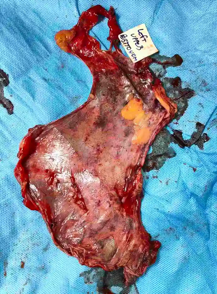
Final histopathology demonstrated residual viable high-grade serous carcinoma involving both ovaries, pelvic peritoneum, uterine serosa, cervix, omentum, bilateral upper peritoneum, appendix, small bowel mesenteric deposits and lesser omentum. The pathological Chemotherapy Response Score (CRS) was 2, indicating a moderate response with substantial residual viable tumour remaining after chemotherapy.
This illustrates an important principle in ovarian cancer management. Chemotherapy may destroy a significant proportion of tumour cells and convert bulky deposits into fibrotic or necrotic lesions, but microscopic viable cancer often persists within these residual deposits. Surgical removal remains essential to achieve maximal disease clearance.
- Clinical Insight
A good response on imaging does not necessarily mean complete eradication of cancer. Residual “burnt-out” deposits may still contain viable tumour and therefore require surgical excision.
Indian Ovarian Cancer Institute (IOCI), Ahmedabad
Every patient is unique. Treatment recommendations are individualized based on clinical evaluation, imaging, pathology and multidisciplinary discussion.
What Does a Chemotherapy Response Score (CRS) Tell Us After Interval Surgery?
⦿ Histopathological examination after interval cytoreductive surgery provides valuable information beyond confirming the diagnosis. In this case, pathology showed residual viable high-grade serous carcinoma involving both ovaries with tumour deposits across multiple peritoneal surfaces, including the pelvic peritoneum, omentum, bilateral upper peritoneum, appendix, small bowel mesentery and lesser omentum. Five of twenty-eight dissected lymph nodes also contained metastatic disease without extranodal extension.
⦿ The specimen received a Chemotherapy Response Score (CRS) 2, indicating a moderate pathological response to neoadjuvant chemotherapy. This means chemotherapy produced significant tumour regression but did not completely eliminate viable cancer cells. CRS is assessed by the pathologist after interval surgery and helps evaluate how effectively the tumour responded to preoperative chemotherapy.
⦿ Unlike CT or PET scans, which assess radiological response, the CRS reflects the actual microscopic effect of chemotherapy on the tumour, providing additional prognostic information and helping guide subsequent adjuvant treatment planning.
- Clinical Insight
Imaging demonstrates how the disease appears after chemotherapy, whereas the Chemotherapy Response Score reveals how much viable cancer actually remains. Both are important in planning comprehensive ovarian cancer treatment.
Indian Ovarian Cancer Institute (IOCI), Ahmedabad
Every patient is unique. Treatment recommendations are individualized based on clinical evaluation, imaging, pathology and multidisciplinary discussion.
Get health updates from Dr Swati Shah on WhatsApp
Swasth Parivar: twice a month, practical advice on recovery, diet and warning signs. No advertising. Stop any time.
Your number is used for these updates and for the clinic to reach you. It is never sold or passed on. To stop, reply STOP on WhatsApp.