


Case Library - CRS - HIPEC
Advanced High-Grade Serous Ovarian Cancer After Neoadjuvant Chemotherapy
⦿ Advanced ovarian cancer often spreads beyond the ovaries before diagnosis, involving the lining of the abdomen (peritoneum), omentum and nearby tissues. In one recent clinical case managed at the Indian Ovarian Cancer Institute (IOCI), Ahmedabad, the patient had high-grade serous ovarian carcinoma involving both ovaries with documented peritoneal and omental disease after receiving three cycles of neoadjuvant chemotherapy. Surgical and histopathology findings confirmed residual tumour involving both ovaries, pelvic peritoneum, cul-de-sac, greater omentum and selected peritoneal deposits, while some other organs remained free of tumour.
⦿ Management included interval cytoreductive surgery followed by HIPEC, aiming to remove all visible disease (CC-0 cytoreduction). The procedure also included removal of involved peritoneal surfaces, omentum and regional lymph node assessment as part of comprehensive treatment planning.
⦿ Clinical Insight: High-grade serous ovarian cancer frequently spreads within the abdominal cavity rather than remaining confined to the ovaries. Careful surgical assessment after chemotherapy plays an important role in determining the extent of residual disease and guiding further treatment.
⦿ This case reflects the multidisciplinary management of advanced ovarian cancer at Indian Ovarian Cancer Institute (IOCI), Ahmedabad.
⦿ Every patient is unique. Treatment recommendations are individualized based on clinical evaluation, imaging, pathology and multidisciplinary discussion.
Why Interval Cytoreductive Surgery Was Performed After Chemotherapy
⦿ Not every patient with advanced ovarian cancer undergoes surgery immediately after diagnosis. In this case, the patient first received three cycles of neoadjuvant chemotherapy with Carboplatin and Paclitaxel before undergoing planned interval cytoreductive surgery. Follow-up imaging documented reduction in the size of both adnexal masses compared with the initial scans, after which surgery was scheduled.
⦿ During surgery, disease involving the ovaries, pelvic peritoneum, omentum and other abdominal surfaces was addressed through comprehensive cytoreduction followed by HIPEC. Histopathology later demonstrated residual viable high-grade serous carcinoma with a Chemotherapy Response Score (CRS2), indicating a moderate pathological response to preoperative chemotherapy.
⦿ Clinical Insight: In selected patients with advanced ovarian cancer, neoadjuvant chemotherapy can reduce tumour burden and may improve the feasibility of achieving optimal cytoreductive surgery. The final decision depends on imaging findings, overall fitness, disease distribution and multidisciplinary evaluation.
⦿ This clinical experience is shared by Indian Ovarian Cancer Institute (IOCI), Ahmedabad.
⦿ Every patient is unique. Treatment recommendations are individualized based on clinical evaluation, imaging, pathology and multidisciplinary discussion.
Understanding Cytoreductive Surgery with HIPEC in Advanced Ovarian Cancer
⦿ Some patients with advanced ovarian cancer require extensive surgery because the disease has spread beyond the ovaries to the lining of the abdomen. In this case, the patient underwent complete cytoreductive surgery (CC-0) combined with Hyperthermic Intraperitoneal Chemotherapy (HIPEC).
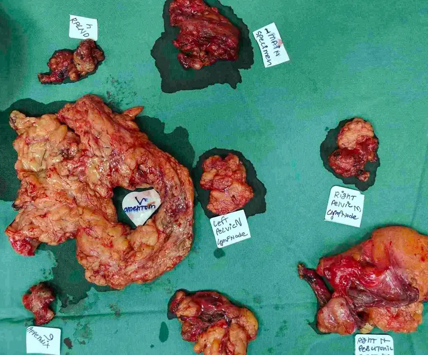
⦿ The operation included removal of both ovaries, uterus, pelvic peritoneum, cul-de-sac, greater and lesser omentum, involved peritoneal surfaces, regional pelvic and para-aortic lymph nodes, mesenteric deposits and umbilicus, followed by HIPEC using heated cisplatin administered within the abdominal cavity. According to the operative record, complete macroscopic cytoreduction (CC-0) was achieved before HIPEC was performed.
⦿ Clinical Insight: Cytoreductive surgery aims to remove all visible tumour deposits whenever feasible. HIPEC is delivered immediately after surgery while the abdomen is still open, allowing heated chemotherapy to circulate within the abdominal cavity. Whether HIPEC is appropriate depends on individual clinical circumstances and multidisciplinary decision-making.
⦿ This case represents one example of advanced ovarian cancer management at Indian Ovarian Cancer Institute (IOCI), Ahmedabad.
⦿ Every patient is unique. Treatment recommendations are individualized based on clinical evaluation, imaging, pathology and multidisciplinary discussion.
What Does a Chemotherapy Response Score (CRS2) Mean?
⦿ After surgery for advanced ovarian cancer, histopathology provides valuable information about how the tumour responded to treatment. In this case, the pathology report showed residual viable high-grade serous carcinoma involving both ovaries with additional tumour deposits in the greater omentum, pelvic peritoneum, cul-de-sac, sigmoid deposit and upper peritoneal biopsies. One of seventeen left pelvic lymph nodes contained metastatic carcinoma, while the remaining pelvic and para-aortic lymph nodes examined were negative. The pathology also assigned a Chemotherapy Response Score (CRS2) for the omentum and ovary.
⦿ A CRS2 score indicates a moderate pathological response to neoadjuvant chemotherapy. This means that chemotherapy produced measurable treatment effect, but viable tumour was still present. The pathology findings help the multidisciplinary team understand disease response and assist in planning subsequent management.
⦿ Clinical Insight: Histopathology does much more than confirm the diagnosis. It evaluates residual disease, treatment response and lymph node involvement, all of which contribute to individualized treatment planning.
⦿ This educational case is shared by Indian Ovarian Cancer Institute (IOCI), Ahmedabad.
⦿ Every patient is unique. Treatment recommendations are individualized based on clinical evaluation, imaging, pathology and multidisciplinary discussion.
Can Ovarian Cancer Present as an Inguinal Lump? The Importance of Lymph Node Biopsy and IHC
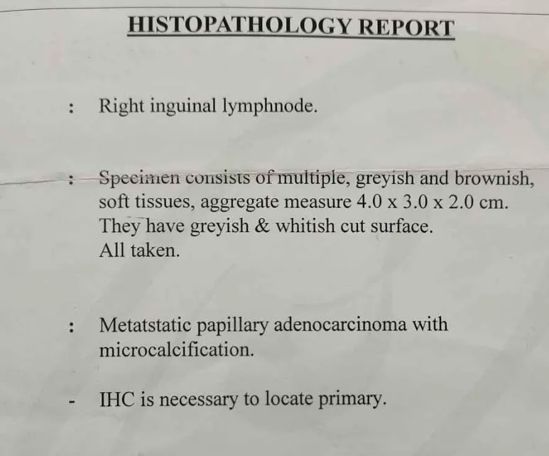
Recently, our team at the Indian Ovarian Cancer Institute (IOCI), Ahmedabad managed an unusual presentation of ovarian cancer. The patient’s first confirmed diagnosis came from an excision biopsy of an enlarged right inguinal lymph node, which demonstrated metastatic papillary adenocarcinoma. Immunohistochemistry (IHC) helped identify the likely ovarian origin, following which further evaluation confirmed ovarian cancer. The patient subsequently received neoadjuvant chemotherapy before undergoing interval cytoreductive surgery.
Many cancers can spread to inguinal lymph nodes, making IHC an important investigation when the primary tumour is not immediately obvious. Rather than treating only the enlarged lymph node, identifying the site of origin allows disease-specific treatment planning.
- Expert Insight
Persistent or unexplained enlarged inguinal lymph nodes should always be evaluated carefully. In selected patients, biopsy and immunohistochemistry can identify the primary cancer and guide appropriate treatment.
Every patient is unique. Treatment recommendations are individualized based on clinical evaluation, imaging, pathology and multidisciplinary discussion.
Can Some Patients with Stage IV Ovarian Cancer Undergo Complete Tumour Removal?
Recently, our team at the Indian Ovarian Cancer Institute (IOCI), Ahmedabad managed a patient with Stage IV ovarian cancer who initially had biopsy-proven inguinal lymph node metastasis. After three cycles of Paclitaxel and Carboplatin chemotherapy, the patient’s CA-125 decreased from 170 U/mL to 11.1 U/mL, and CT imaging demonstrated a marked reduction in disease with disappearance of previously enlarged lymph nodes.
Following reassessment, the patient underwent CC-0 cytoreductive surgery, meaning no visible tumour remained at the completion of surgery. Comprehensive surgery included hysterectomy, bilateral salpingo-oophorectomy, pelvic peritonectomy, pelvic and retroperitoneal lymph node dissection, and supracolic omentectomy.
- Clinical Pearl
Not every Stage IV ovarian cancer behaves the same way. In carefully selected patients who respond well to chemotherapy, complete cytoreduction may become achievable as part of multidisciplinary treatment planning.
Every patient is unique. Treatment recommendations are individualized based on clinical evaluation, imaging, pathology and multidisciplinary discussion.
I recommend using “undergo complete tumour removal” rather than “can be cured.” The documents show CC-0 surgery but do not establish cure.
Why Complete Surgery Is Performed Even When the Ovaries Appear Almost Normal
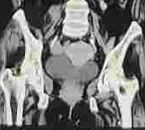
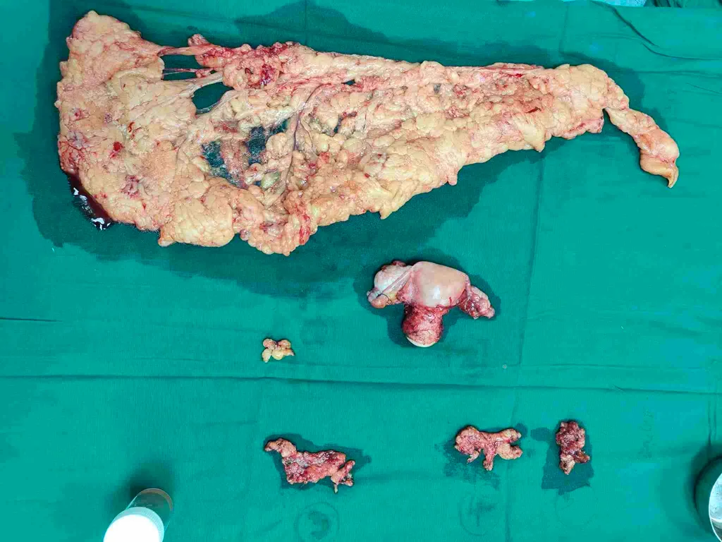
Recently, our team at the Indian Ovarian Cancer Institute (IOCI), Ahmedabad managed a patient whose ovaries appeared grossly normal during surgery after neoadjuvant chemotherapy. Apart from small deposits near the fimbrial ends of the fallopian tubes and mildly enlarged pelvic and retroperitoneal lymph nodes, there was no obvious extensive abdominal disease.
Despite this reassuring appearance, comprehensive cytoreductive surgery was performed. Histopathological examination later confirmed residual viable high-grade serous carcinoma in the right ovary, while the left ovary, omentum, upper peritoneum and all 27 dissected lymph nodes were free of tumour.
- What Patients Should Know:
Chemotherapy can significantly alter the appearance of ovarian cancer. Even when organs look almost normal during surgery, microscopic cancer may still be present. Histopathology remains the definitive method for assessing residual disease.
Every patient is unique. Treatment recommendations are individualized based on clinical evaluation, imaging, pathology and multidisciplinary discussion.
Does a Good CT Scan Mean All the Cancer Has Gone?
Recently, our team at the Indian Ovarian Cancer Institute (IOCI), Ahmedabad managed a patient whose CT scan after chemotherapy showed a marked reduction in the ovarian tumour, disappearance of previously enlarged lymph nodes and normalization of CA-125. These findings indicated an excellent clinical response before surgery.
However, the final histopathology told a more detailed story. Examination of the surgical specimen confirmed residual viable high-grade serous carcinoma in the right ovary. The pathology also assigned a Chemotherapy Response Score (CRS1), indicating limited pathological response to chemotherapy, although the omentum, upper peritoneum and all regional lymph nodes were free of tumour.
Did You Know? Imaging and tumour markers are valuable tools for monitoring treatment response, but only histopathological examination can determine the amount of residual viable cancer after surgery.
Every patient is unique. Treatment recommendations are individualized based on clinical evaluation, imaging, pathology and multidisciplinary discussion.
Why Do Some Patients with Advanced Ovarian Cancer Receive Chemotherapy Before Surgery?
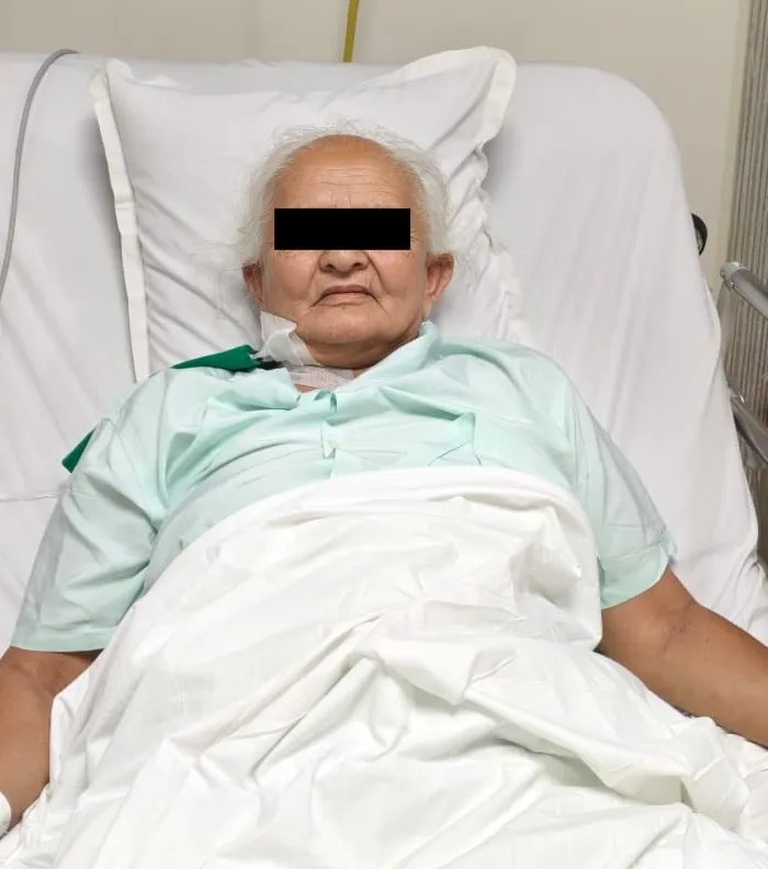
Not every patient with advanced ovarian cancer benefits from immediate surgery. In one recently managed case at the Indian Ovarian Cancer Institute (IOCI), Ahmedabad, the patient presented with subacute intestinal obstruction, extensive peritoneal disease and reduced performance status. Rather than proceeding directly to major surgery, the multidisciplinary team recommended neoadjuvant chemotherapy first.
After three cycles of Carboplatin and Paclitaxel, the patient’s condition improved remarkably. CT imaging showed significant reduction in disease burden, ascites resolved and the CA-125 level reduced from 421 U/mL to 22.6 U/mL, making interval cytoreductive surgery feasible.
- Expert Insight
The decision between Primary Cytoreductive Surgery (PCS) and Interval Cytoreductive Surgery (ICS) is one of the most important decisions in ovarian cancer treatment. It depends on disease distribution, the likelihood of complete tumour removal, the patient’s nutritional status, overall fitness and expected surgical safety—not simply the stage of the disease.
Choosing the right treatment sequence can improve the chances of successful surgery while reducing treatment-related risks.
Can Ovarian Cancer Attached to the Bowel Be Removed Without Removing the Bowel?
Patients are often worried that ovarian cancer involving the bowel always requires bowel resection. However, this is not true in every case.
During interval cytoreductive surgery in one recently managed patient at the Indian Ovarian Cancer Institute (IOCI), Ahmedabad, tumour deposits were found close to the transverse colon, sigmoid colon and upper rectum. Careful surgical assessment showed that the disease involved the outer surface (serosa) rather than deeply invading the bowel wall. This allowed complete tumour clearance while preserving the bowel, and CC-0 cytoreduction (no visible residual disease) was achieved.
- Surgical Perspective
The decision to remove a segment of bowel is based on the depth of tumour invasion, not simply because the tumour is touching the bowel. In carefully selected patients, experienced ovarian cancer surgeons may safely preserve the bowel while still achieving complete cytoreduction.
Organ preservation, whenever oncologically appropriate, can reduce surgical complexity without compromising cancer surgery principles.
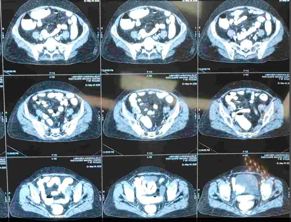
What Is the Peritoneal Cancer Index (PCI) and Why Is It Important in Ovarian Cancer Surgery?
The Peritoneal Cancer Index (PCI) is a scoring system used to estimate how extensively ovarian cancer has spread within the abdominal cavity. It helps surgeons understand disease distribution and plan cytoreductive surgery.
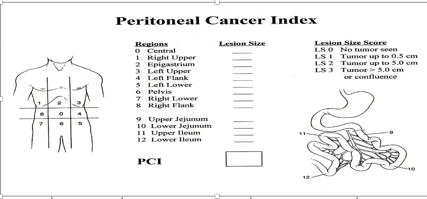
In one recently treated patient at the Indian Ovarian Cancer Institute (IOCI), Ahmedabad, the PCI was 13, indicating significant peritoneal disease. Despite this, comprehensive interval cytoreductive surgery was successfully performed. After removal of all visible tumour deposits, the surgery achieved CC-0 cytoreduction, meaning no macroscopic residual disease remained.
- Did You Know?
A higher PCI score does not automatically mean surgery is impossible. The final decision depends on whether complete tumour removal appears achievable while maintaining patient safety. Disease location, organ involvement, response to chemotherapy and surgical expertise all influence treatment planning.
How Does Histopathology Show the Response to Chemotherapy in Ovarian Cancer?
After chemotherapy and surgery, the most accurate assessment of treatment response comes from histopathological examination.
In this recently managed case at the Indian Ovarian Cancer Institute (IOCI), Ahmedabad, pathology confirmed residual high-grade serous carcinoma involving the left ovary, pelvic peritoneum, omentum, appendix serosa and small paracolic peritoneal deposits. The report assigned a Chemotherapy Response Score (CRS2) for both the left adnexa and omentum, indicating a moderate response to neoadjuvant chemotherapy. Four of thirty-four lymph nodes showed metastatic disease, while the umbilicus was free of tumour.
- Understanding the Report
CT scans and tumour markers help assess treatment response, but they cannot determine the exact amount of residual viable cancer. Histopathology provides this information and plays a vital role in confirming disease extent, evaluating chemotherapy response and planning further treatment.
Each pathology report adds an important piece to the overall treatment strategy.
Bilateral Adnexal Mass + Omental Deposits + Peritoneal Carcinomatosis: Why Biopsy with IHC Is Essential Before Starting Chemotherapy
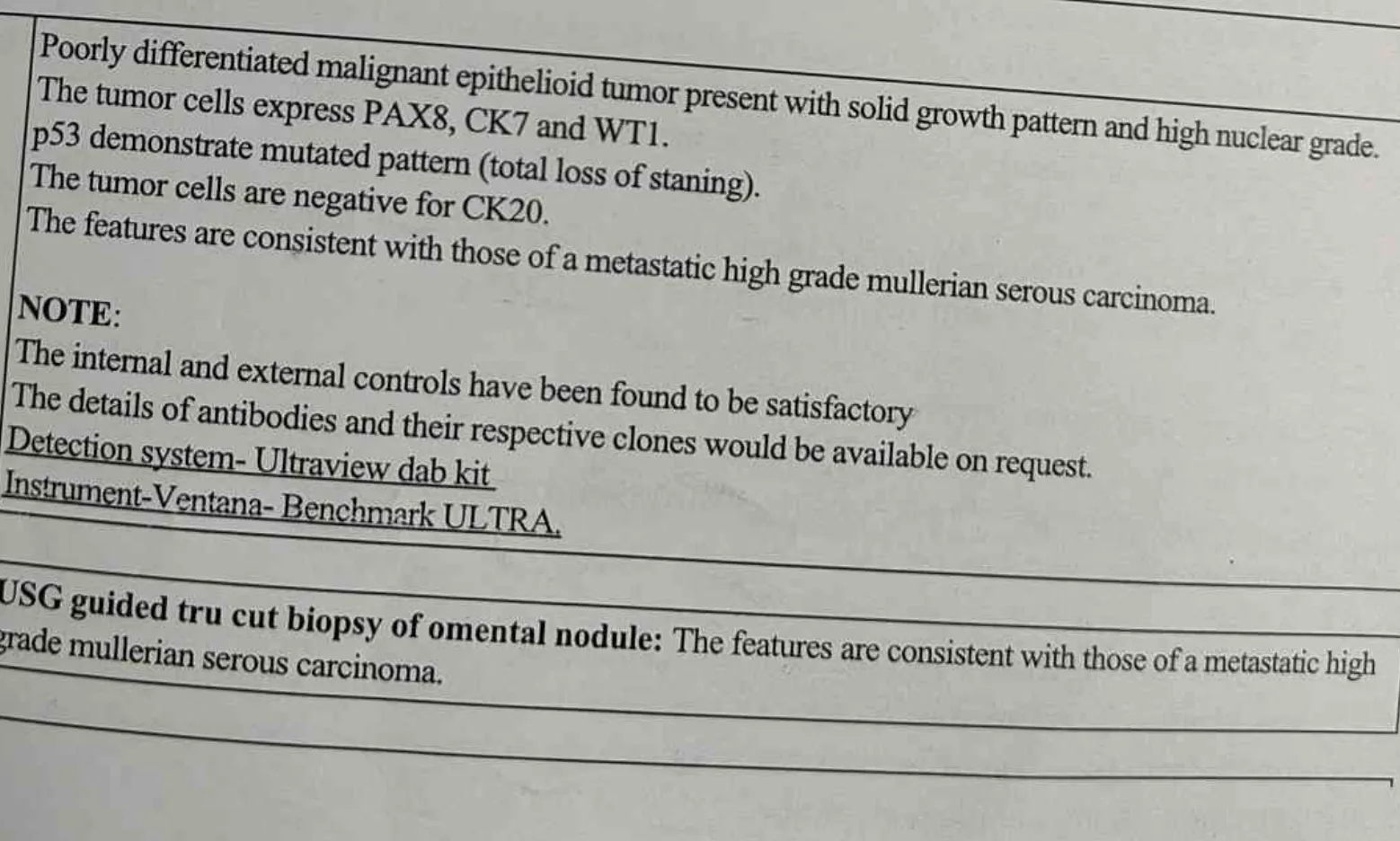
A recently managed case at the Indian Ovarian Cancer Institute (IOCI), Ahmedabad presented with bilateral adnexal masses, multiple omental and peritoneal deposits, mesenteric deposits and peritoneal carcinomatosis. An USG-guided tru-cut biopsy of the omental nodule followed by Immunohistochemistry (IHC) confirmed metastatic high-grade Müllerian serous carcinoma, establishing the diagnosis before neoadjuvant chemotherapy was started.
- Why is this important?
- When scans show:
⦿ Bilateral ovarian / adnexal masses
⦿ Omental deposits
⦿ Peritoneal deposits
⦿ Mesenteric deposits
⦿ Ascites
⦿ Peritoneal carcinomatosis
⦿ the primary cancer may occasionally arise from the ovary, fallopian tube, endometrium, stomach, colon, pancreas or biliary tract.
⦿ An image-guided biopsy with Immunohistochemistry (IHC) confirms the tissue of origin and helps select the correct chemotherapy protocol.
Every patient is unique. Treatment recommendations are individualized based on clinical evaluation, imaging, pathology and multidisciplinary discussion.
I recommend using “undergo complete tumour removal” rather than “can be cured.” The documents show CC-0 surgery but do not establish cure.
Sigmoid Colon, Rectum or Bowel Involvement in Ovarian Cancer: Can Complete Cytoreductive Surgery Still Be Performed?
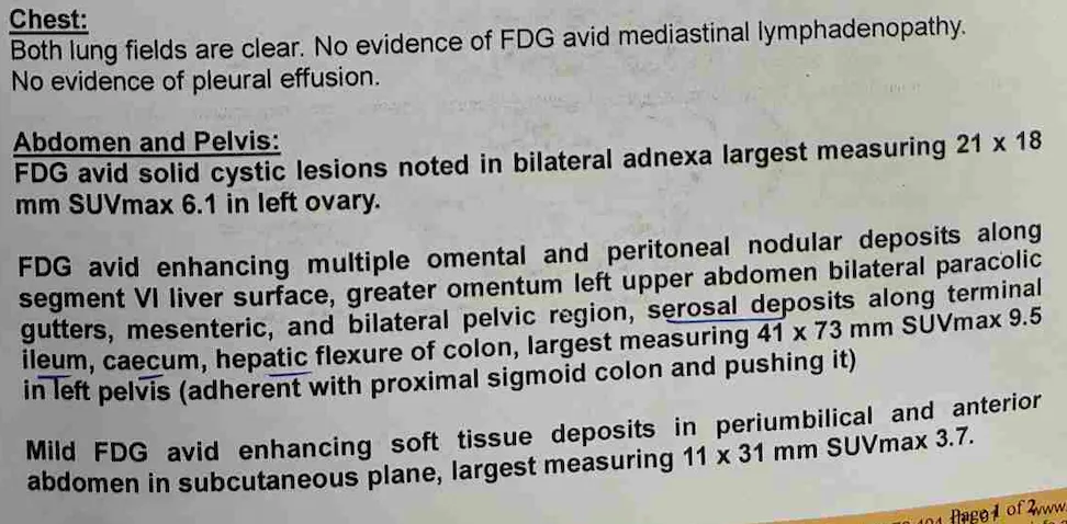
A recently operated case at the Indian Ovarian Cancer Institute (IOCI), Ahmedabad had peritoneal carcinomatosis with tumour deposits involving the sigmoid colon, rectum, terminal ileum, mesentery and right colon. Complete treatment included Low Anterior Resection (LAR), Right Hemicolectomy, bowel anastomosis, Cytoreductive Surgery (CRS) and HIPEC.
- Clinical Insight
- Many PET-CT reports mention:
⦿ Sigmoid colon involvement
⦿ Rectal involvement
⦿ Serosal bowel deposits
⦿ Mesenteric deposits
⦿ Bowel wall involvement
⦿ These findings do not automatically mean surgery is impossible.
- Modern Cytoreductive Surgery (CRS) may include:
⦿ Low Anterior Resection (LAR)
⦿ Right Hemicolectomy
⦿ Small bowel resection (when required)
⦿ Safe bowel anastomosis
⦿ Complete removal of visible peritoneal disease
A temporary ileostomy or colostomy may occasionally be required to protect the bowel anastomosis.
In most ovarian cancer patients, if a stoma is needed, it is temporary rather than permanent and may be reversed after healing.
Take-home message:
Even when ovarian cancer involves the bowel, complete cytoreductive surgery with bowel reconstruction is often possible in carefully selected patients.
Peritoneal Carcinomatosis in Ovarian Cancer: Why HIPEC Targets Surface Disease
A recently operated case at the Indian Ovarian Cancer Institute (IOCI), Ahmedabad showed multiple omental deposits, peritoneal deposits, mesenteric deposits, liver capsule deposits, falciform ligament deposits, pelvic peritoneal deposits and bowel serosal deposits, followed by Cytoreductive Surgery (CRS) with HIPEC.

- Clinical Insight
- Advanced ovarian cancer commonly spreads over the peritoneal surfaces , producing:
⦿ Peritoneal carcinomatosis
⦿ Omental caking
⦿ Mesenteric deposits
⦿ Pelvic peritoneal deposits
⦿ Diaphragmatic peritoneal deposits
⦿ Liver capsule deposits
⦿ Bowel serosal deposits
Unlike cancers that primarily spread into solid organs, ovarian cancer often spreads across the surface lining of the abdomen (peritoneum).
After complete Cytoreductive Surgery (CRS), HIPEC (Hyperthermic Intraperitoneal Chemotherapy) delivers heated chemotherapy directly inside the abdominal cavity to treat microscopic residual disease present on these peritoneal surfaces.
Take-home message:
The biology of peritoneal surface spread is one of the reasons why CRS combined with HIPEC may benefit selected patients with advanced ovarian cancer.
Recovery After Extensive Cytoreductive Surgery (CRS) and HIPEC for Advanced Ovarian Cancer
A recently operated case at the Indian Ovarian Cancer Institute (IOCI), Ahmedabad underwent 11-hour Cytoreductive Surgery with HIPEC, for advanced ovarian cancer with peritoneal carcinomatosis.
- Clinical Insight
- Patients often search:
⦿ Recovery after ovarian cancer surgery
⦿ Recovery after HIPEC
⦿ Recovery after bowel resection
⦿ Recovery after CRS surgery
⦿ How long to recover after ovarian cancer operation
- Although these procedures are extensive, recovery has improved because of:
⦿ ERAS (Enhanced Recovery After Surgery) protocols
⦿ Better anaesthesia
⦿ Early mobilisation
⦿ Early oral nutrition
⦿ Effective pain management
⦿ Physiotherapy
⦿ Multidisciplinary perioperative care
⦿ Strong family support and patient motivation
Many patients gradually return to walking, eating, daily activities and further chemotherapy within a planned recovery pathway.
Take-home message:
The extent of surgery alone should not determine treatment decisions. With experienced Cytoreductive Surgery (CRS), HIPEC, ERAS protocols and multidisciplinary care, recovery after advanced ovarian cancer surgery can be faster and smoother than many patients expect.
Retroperitoneal Lymph Nodes (RPLN) in Ovarian Cancer: Does It Always Mean Chemotherapy First?
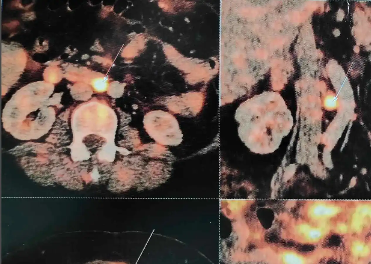
A recently operated case at the Indian Ovarian Cancer Institute (IOCI), Ahmedabad had bilateral ovarian masses, diffuse omental metastasis and FDG-avid aortocaval (retroperitoneal) lymph nodes on PET-CT.
- Many patients believe:
“If retroperitoneal lymph nodes (RPLN) are involved, surgery cannot be done.”
This is not true in ovarian cancer.
Unlike many other cancers, retroperitoneal lymph node involvement alone does NOT make ovarian cancer unresectable.
- The most important question is:
Can Complete Cytoreductive Surgery (CC0) be achieved?
That decision should be made only after evaluation by an experienced Gynecologic Oncologist / Ovarian Cancer Surgeon / Peritoneal Surface Malignancy Surgeon.
If complete tumour clearance appears achievable and the patient is fit, Primary Cytoreductive Surgery may still be the best first treatment—even in the presence of enlarged pelvic or retroperitoneal lymph nodes.
Take-home message:
In ovarian cancer, RPLN involvement is not an automatic indication for Neoadjuvant Chemotherapy. The treatment sequence should be individualized after expert surgical assessment.
Diagnostic Laparoscopy in Ovarian Cancer: Confirming Resectability Before Major Surgery
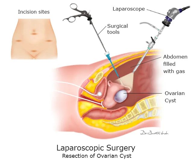
A recently managed case at the Indian Ovarian Cancer Institute (IOCI), Ahmedabad underwent Staging Diagnostic Laparoscopy before definitive surgery.
- Laparoscopy demonstrated:
✔ Bilateral adnexal masses
✔ Approximately 300 ml free fluid
✔ Omental caking adherent to the anterior abdominal wall
✔ Small peritoneal deposits over the upper abdomen and terminal ileal mesentery
During the same procedure, peritoneal biopsy and peritoneal fluid sampling were performed.
Most importantly, the extent of peritoneal disease was assessed and the Peritoneal Cancer Index (PCI) was calculated during surgery.
As complete tumour clearance appeared achievable and the patient was fit for major surgery, the team proceeded with Primary CRS + HIPEC, achieving CC0 Cytoreduction.
Take-home message:
Diagnostic laparoscopy is not done simply to “look inside.” It helps assess resectability, PCI, distribution of peritoneal disease, and suitability for Primary Cytoreductive Surgery versus Neoadjuvant Chemotherapy.
Why Biopsy and IHC Are Essential Before Starting Treatment for Advanced Ovarian Cancer
A recently managed case at the Indian Ovarian Cancer Institute (IOCI), Ahmedabad presented with:
⦿ Bilateral ovarian masses
⦿ Diffuse omental metastasis
⦿ Peritoneal carcinomatosis
⦿ Enlarged pelvic and retroperitoneal lymph nodes on PET-CT.
⦿ Before starting definitive treatment, Diagnostic Laparoscopy with Peritoneal Biopsy was performed.
⦿ Histopathology confirmed Metastatic Carcinoma, most likely High-Grade Serous Carcinoma of Ovarian Origin.
⦿ In patients with peritoneal carcinomatosis, omental deposits or peritoneal nodules, tissue diagnosis is extremely important.
Biopsy with Histopathology and Immunohistochemistry (IHC) helps:
✔ Confirm malignancy
✔ Confirm ovarian origin
✔ Exclude metastasis from gastrointestinal or other primary cancers
✔ Plan the correct sequence of treatment
At the same time, diagnostic laparoscopy allows assessment of resectability before committing the patient to major Cytoreductive Surgery.
Take-home message:
For advanced ovarian cancer with peritoneal disease, Diagnostic Laparoscopy + Biopsy + IHC provides both diagnostic confirmation and surgical decision-making, helping deliver the most appropriate treatment.
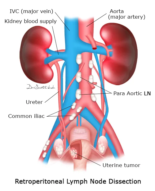
Retroperitoneal Lymph Node Dissection (RPLND) in Ovarian Cancer: Why Experience Matters
A recently operated case at the Indian Ovarian Cancer Institute (IOCI), Ahmedabad underwent Complete Cytoreductive Surgery (CC0) with Pelvic Lymph Node Dissection and Retroperitoneal Lymph Node Dissection (RPLND) as part of comprehensive ovarian cancer surgery.
- Many patients are anxious when imaging mentions:
“Aortocaval lymph node” or “Retroperitoneal lymph node.”
The ovaries naturally drain to the pelvic and para-aortic (retroperitoneal) lymph nodes, making these nodal basins important sites of disease spread.
RPLND is a technically demanding procedure because these lymph nodes lie close to major blood vessels such as the aorta, inferior vena cava and their branches.
However, when indicated and performed by an experienced Gynecologic Oncology team familiar with retroperitoneal anatomy, Retroperitoneal Lymph Node Dissection can be performed safely and meticulously as part of complete cytoreductive surgery.
Take-home message:
Retroperitoneal lymph node involvement in ovarian cancer should not be viewed as “inoperable.” Careful assessment and meticulous surgery by an experienced ovarian cancer surgeon can allow complete treatment in appropriately selected patients.
Advanced Stage IIIC Ovarian Cancer Does Not Always Need Chemotherapy First
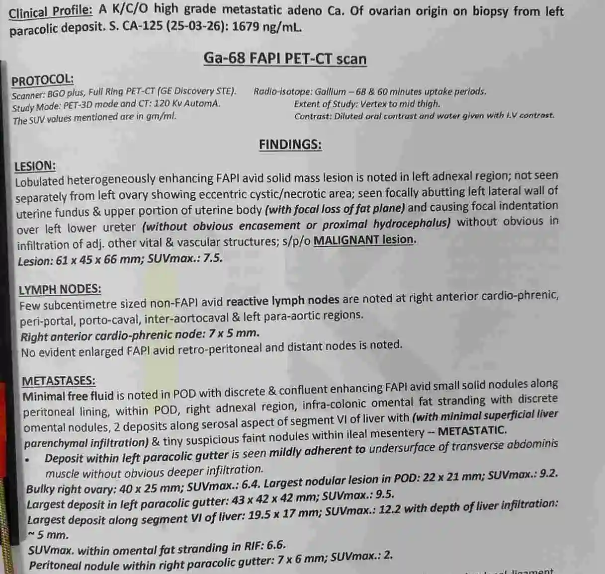
A 46-year-old woman presented with biopsy-proven high-grade serous ovarian carcinoma and a CA-125 of 1679 U/mL. Ga-68 FAPI PET-CT demonstrated a left adnexal malignancy with peritoneal deposits involving the pouch of Douglas (POD), omentum, right adnexal region, left paracolic gutter, superficial deposits over segment VI of the liver and tiny ileal mesenteric nodules. The Peritoneal Cancer Index (PCI) was reported as 13.
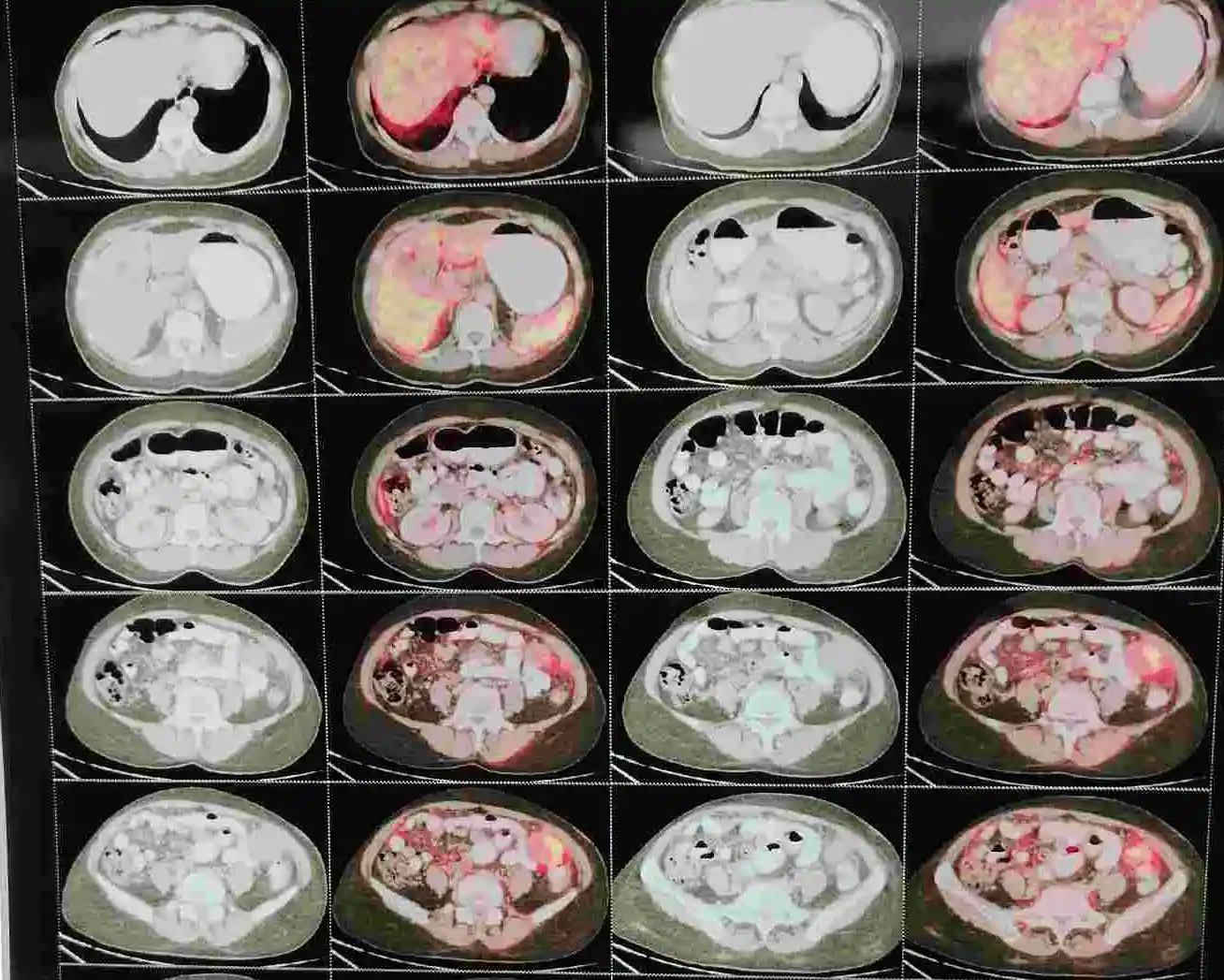
⦿ At first glance, this appears to be advanced FIGO Stage IIIC ovarian cancer. However, the decision between Primary Cytoreductive Surgery (CRS) and Neoadjuvant Chemotherapy (NACT) depends less on stage and more on whether complete tumour removal is realistically achievable.
⦿ In this patient, imaging showed no disease involving the hepatoduodenal ligament, mesenteric root, urinary bladder, serosal surface of both ureters, splenic surface or left hemidiaphragm, and there were no enlarged FAPI-avid retroperitoneal or distant lymph nodes. These anatomical locations are important because extensive tumour at these sites may reduce the likelihood of complete cytoreduction.
⦿ A staging laparoscopy was therefore performed first, confirming operability before proceeding to complete cytoreductive surgery with HIPEC.
- Clinical Insight
⦿ In advanced ovarian cancer, the critical question is not simply “What is the stage?” but “Can all visible disease be safely removed?” Careful analysis of disease distribution often determines the initial treatment strategy.
⦿ This case reflects real-world surgical decision-making at the Indian Ovarian Cancer Institute (IOCI), Ahmedabad.
⦿ Every patient is unique. Treatment recommendations are individualized based on clinical evaluation, imaging, pathology and multidisciplinary discussion.
Take-home message:
The biology of peritoneal surface spread is one of the reasons why CRS combined with HIPEC may benefit selected patients with advanced ovarian cancer.
How Ga-68 FAPI PET-CT Helped Map Peritoneal Disease Before Ovarian Cancer Surgery
⦿ This patient underwent Ga-68 FAPI PET-CT, which provided detailed mapping of tumour distribution before surgery. Besides identifying the primary left ovarian mass, the scan demonstrated metastatic deposits along the pelvic peritoneum, pouch of Douglas, omentum, right adnexa, left paracolic gutter, segment VI liver capsule and tiny ileal mesenteric nodules. Equally important was what the scan did not show.
⦿ There were no FAPI-avid deposits involving the hepatoduodenal ligament, mesenteric root, undersurface of the left hemidiaphragm, urinary bladder, serosal surface of the ureters or spleen. No enlarged FAPI-avid retroperitoneal or distant lymph nodes were identified. The reported PCI was 13/39.
⦿ For surgeons planning cytoreductive surgery, this information is valuable because it estimates the extent of peritoneal disease and identifies surgically challenging anatomical regions before entering the operating room.
⦿ During surgery, the operative findings closely correlated with the imaging, and complete cytoreduction was achieved before HIPEC.
- Clinical Insight
Advanced imaging should not only detect ovarian cancer—it should also define where the disease is and where it is not, allowing more informed surgical planning.
This reflects the practical use of advanced imaging at the Indian Ovarian Cancer Institute (IOCI), Ahmedabad.
Every patient is unique. Treatment recommendations are individualized based on clinical evaluation, imaging, pathology and multidisciplinary discussion.
Liver Deposits in Ovarian Cancer Do Not Always Mean the Liver Needs to Be Removed
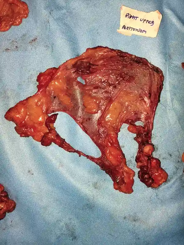
One of the important findings in this patient was tumour adjacent to segment VI of the liver. Imaging described minimal superficial liver infiltration, while the operative findings confirmed that the deposits were adherent to the liver surface without parenchymal extension. This distinction is clinically important.
Ovarian cancer most commonly spreads over the peritoneal surfaces covering abdominal organs rather than growing deep into the organs themselves. In this patient, the liver deposits involved the capsule (surface) rather than liver tissue. Similarly, the lesion described in the left paracolic gutter represented disease adherent to the peritoneal and omental surfaces rather than deep invasion of the abdominal wall.
Recognising this pattern allowed complete removal of visible disease while preserving the liver itself. The operative note also documented that the liver was otherwise grossly normal, with no parenchymal tumour extension.
- Clinical Insight
In ovarian cancer, the location and depth of tumour involvement are often more important than the presence of a deposit alone. Careful assessment helps determine whether organ-preserving cytoreductive surgery is feasible.
This case illustrates this principle at the Indian Ovarian Cancer Institute (IOCI), Ahmedabad.
Every patient is unique. Treatment recommendations are individualized based on clinical evaluation, imaging, pathology and multidisciplinary discussion.
Why HIPEC Was Performed After Complete Cytoreductive Surgery in This Patient
Following complete cytoreductive surgery, this patient underwent Hyperthermic Intraperitoneal Chemotherapy (HIPEC) using cisplatin 150 mg in 4 litres of dialysate at 41°C for 90 minutes.
⦿ The rationale was closely related to the pattern of disease documented before surgery. Imaging and operative findings demonstrated multiple tumour deposits over the pelvic peritoneum, omentum, paracolic gutters and upper abdominal peritoneal surfaces, which are characteristic sites of peritoneal spread in ovarian cancer.
⦿ After all visible tumour deposits had been surgically removed, HIPEC was administered within the abdominal cavity before closure. This approach aims to expose any remaining microscopic peritoneal tumour cells to heated intraperitoneal chemotherapy immediately after cytoreduction.
⦿ HIPEC was used after complete tumour removal—not as a substitute for surgery. Its role was considered only after staging laparoscopy confirmed operability and complete cytoreduction had been achieved.
- Clinical Insight
HIPEC is most appropriately considered in selected patients where extensive peritoneal disease has been surgically cleared. The effectiveness of HIPEC is closely linked to the quality of cytoreductive surgery.
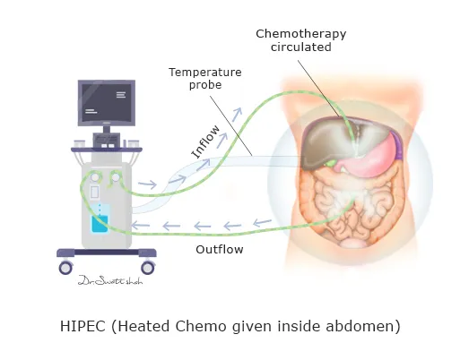
This case demonstrates multidisciplinary management at the Indian Ovarian Cancer Institute (IOCI), Ahmedabad.
Every patient is unique. Treatment recommendations are individualized based on clinical evaluation, imaging, pathology and multidisciplinary discussion.
When Omental and Peritoneal Nodules Need Tissue Diagnosis
A 66-year-old woman presented with abdominal pain, weakness, loss of appetite and weight loss. CT showed mildly bulky, lobulated ovaries with heterogeneous enhancement, closely related to the uterus and anterior rectal wall. It also described a 27 × 16 mm enhancing soft-tissue density in the right iliac fossa adjacent to the base of the caecum, with the appendix not separately visualized, along with diffuse enhancing soft-tissue nodules involving the omental fat and peritoneal lining, irregular peritoneal thickening and moderate-to-gross ascites.
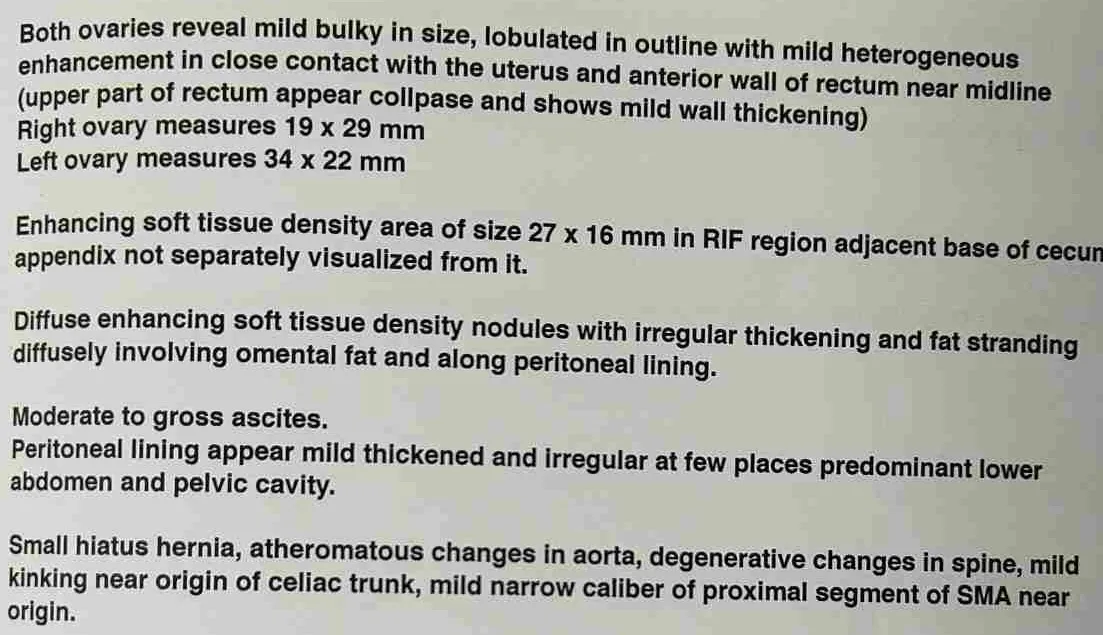
⦿ Such imaging can strongly suggest advanced ovarian malignancy, but the radiological appearance of peritoneal nodules is not, by itself, a substitute for tissue diagnosis. In this patient, the MRI also raised the possibility of a primary appendiceal neoplasm with metastasis.
⦿ A staging laparoscopy with omental biopsy was therefore performed. Importantly, laparoscopy provided direct assessment of the peritoneal cavity: there was gross ascites, pelvic and bilateral paracolic-gutter deposits, bilateral subdiaphragmatic peritoneal deposits, multiple deposits over the whole small-bowel mesentery and omental caking. Three litres of ascitic fluid were aspirated and an omental nodule was sent for histopathology.
⦿ Histology demonstrated infiltrative high-grade carcinoma with solid and tubular patterns, reported as consistent with high-grade serous carcinoma in the appropriate clinical and CA-125 context.
- Clinical Insight
When peritoneal or omental nodules are small or equivocal, image-guided biopsy may not adequately represent the overall disease. Laparoscopy can simultaneously inspect the entire peritoneal cavity, assess disease distribution and obtain a representative biopsy.
Indian Ovarian Cancer Institute (IOCI), Ahmedabad
Every patient is unique. Treatment recommendations are individualized based on clinical evaluation, imaging, pathology and multidisciplinary discussion.
Why Staging Laparoscopy Can Change the Plan in Apparent Stage III Ovarian Cancer
⦿ This patient had imaging showing bilateral bulky, lobulated ovaries, omental and peritoneal nodules, irregular peritoneal thickening and moderate-to-gross ascites, with an additional right iliac-fossa soft-tissue lesion close to the caecal base. MRI suggested bilateral ovarian carcinoma with peritoneal/omental metastases, while also raising the possibility of an appendiceal primary.
⦿ The key surgical question was not simply whether there was ovarian cancer, but whether complete cytoreduction could realistically be achieved.
⦿ Staging laparoscopy provided information that cross-sectional imaging could not fully establish. Operatively, there was gross ascites, pelvic and bilateral paracolic-gutter peritoneal deposits, bilateral subdiaphragmatic deposits, omental caking and multiple deposits distributed over the whole small-bowel mesentery.
⦿ This distribution is particularly important. Extensive disease involving multiple areas of the small-bowel mesentery can make complete macroscopic cytoreduction technically unsafe or unlikely to achieve a CC-0 result, even when CT appears potentially operable.
⦿ The patient therefore proceeded with neoadjuvant chemotherapy, with the treatment strategy subsequently reassessed for cytoreductive surgery and HIPEC.
- Clinical Insight
Staging laparoscopy is not simply a diagnostic procedure. In advanced ovarian cancer, it can function as a surgical feasibility assessment, helping determine whether primary cytoreduction is appropriate or whether chemotherapy should precede surgery. The decision depends on the distribution and resectability of disease, not merely the FIGO stage.
Indian Ovarian Cancer Institute (IOCI), Ahmedabad
Every patient is unique. Treatment recommendations are individualized based on clinical evaluation, imaging, pathology and multidisciplinary discussion.
Bilateral Ovarian Masses: Confirm the Primary Before Starting Treatment
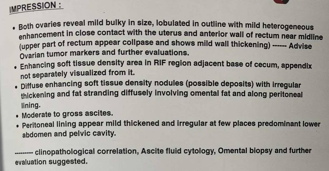
This 66-year-old woman had bilateral ovarian abnormalities on imaging. CT described both ovaries as mildly bulky, lobulated and heterogeneously enhancing, measuring 19 × 29 mm on the right and 34 × 22 mm on the left, with close contact with the uterus and anterior rectal wall. The scan also showed an enhancing lesion near the base of the caecum, with the appendix not separately visualized, together with diffuse omental and peritoneal nodules and moderate-to-gross ascites.
⦿ This pattern can represent a primary ovarian carcinoma, but bilateral ovarian masses should also raise the possibility of metastatic disease. Possible primary sites include the gastrointestinal tract, breast and pancreatobiliary/ hepatobiliary system. In this case, the radiologist specifically mentioned the possibility of a primary appendiceal neoplasm with metastasis.
⦿ Therefore, tissue confirmation is important before assigning the origin of the cancer. Laparoscopy demonstrated extensive peritoneal disease and allowed an omental nodule to be biopsied. Histopathology showed high-grade carcinoma with solid and tubular architecture, considered consistent with high-grade serous carcinoma in the appropriate clinical and CA-125 context.
⦿ When the origin remains uncertain, immunohistochemistry can help establish lineage. A panel may include PAX8/WT1 and ER when Müllerian/ovarian origin is considered, while CK20, CDX2 and SATB2 can support gastrointestinal origin. Depending on the clinical differential, breast markers such as GATA3 and site-specific hepatobiliary markers may also be considered.
- Clinical Insight:
A bilateral ovarian mass is not automatically a primary ovarian cancer. Biopsy plus appropriate IHC can prevent treatment being based on an incorrect assumption about the primary site.
Indian Ovarian Cancer Institute (IOCI), Ahmedabad
Every patient is unique. Treatment recommendations are individualized based on clinical evaluation, imaging, pathology and multidisciplinary discussion.
Staging Laparoscopy Can Quantify Peritoneal Disease Before Major Cytoreductive Surgery
⦿ In advanced ovarian cancer, knowing that peritoneal disease is present is not enough. The distribution and volume of tumour deposits determine whether complete cytoreduction is realistically achievable.
⦿ In this patient, CT demonstrated diffuse enhancing soft-tissue density nodules with irregular thickening and fat stranding involving the omental fat and peritoneal lining, associated with moderate-to-gross ascites. The peritoneal lining was described as mildly thickened and irregular, predominantly in the lower abdomen and pelvis.
⦿ Staging laparoscopy provided a much more direct assessment. It documented gross ascites, pelvic deposits, bilateral paracolic-gutter deposits, bilateral subdiaphragmatic peritoneal deposits, omental caking and multiple deposits over the whole small-bowel mesentery. Three litres of ascitic fluid were aspirated during the procedure.
⦿ This type of laparoscopic assessment can also be used to calculate the Peritoneal Cancer Index (PCI) by systematically assessing tumour involvement across defined abdominal and pelvic regions. PCI provides a structured estimate of peritoneal tumour burden and, particularly when combined with assessment of small-bowel and mesenteric involvement, helps in planning cytoreductive surgery.
⦿ The importance is practical: a patient may appear operable on CT, but direct laparoscopic inspection may reveal disease distribution that makes CC-0 cytoreduction unlikely. Such information can support a decision for neoadjuvant chemotherapy before reassessment for cytoreductive surgery and HIPEC.
- Clinical Insight
In peritoneal malignancy, mapping disease is as important as detecting disease. Laparoscopy can convert a general radiological impression into a surgical assessment of tumour distribution, resectability and cytoreductive feasibility.
Indian Ovarian Cancer Institute (IOCI), Ahmedabad
Every patient is unique. Treatment recommendations are individualized based on clinical evaluation, imaging, pathology and multidisciplinary discussion.
Stage IV Ovarian Cancer: Is Cure Still Possible?
Stage IV ovarian cancer does not automatically mean that treatment is only palliative.
This 51-year-old woman presented with Stage IV high-grade serous ovarian carcinoma, with disease involving the ovaries, peritoneum, omentum and lymph nodes, along with thoracic disease. Her initial CA-125 was 4282.
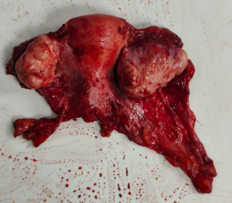
- She received 3 cycles of paclitaxel + carboplatin , with bevacizumab in the first two cycles. Her CA-125 fell dramatically from 4282 → 300 → 157 . The subsequent PET-CT showed:
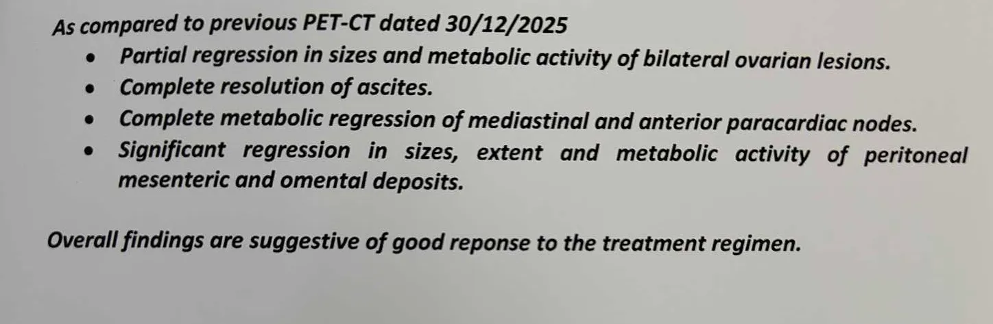
⦿ Complete metabolic resolution of the mediastinal/paracardiac nodes
⦿ Complete resolution of ascites
⦿ Significant regression of peritoneal, mesenteric and omental disease
⦿ Residual disease predominantly within the abdomen/pelvis
⦿ This response made cytoreductive surgery feasible.
⦿ She subsequently underwent extensive cytoreductive surgery (CRS) with HIPEC, including hysterectomy with BSO, pelvic and upper peritonectomy, omentectomy, lymph-node dissection and removal of deposits from the mesentery, bowel surface and hepatoduodenal ligament.
⦿ The operative PCI was 26 before cytoreduction, demonstrating a substantial initial peritoneal tumour burden. Postoperatively, the recorded PCI was 2, reflecting a dramatic reduction in visible peritoneal disease.
- The learning point
Stage IV ovarian cancer can still be approached with an aggressive, potentially long-term disease-control strategy in carefully selected patients.
Response to systemic chemotherapy can convert initially extensive disease into a surgically treatable situation. When complete or near-complete cytoreduction is achievable, CRS ± HIPEC may form an important part of multimodality treatment.
Stage IV is a stage—not a statement that treatment is futile.
Indian Ovarian Cancer Institute (IOCI), Ahmedabad
Every patient is unique. Treatment is individualized according to disease distribution, response to chemotherapy, surgical feasibility and overall patient fitness.
Bevacizumab in Advanced Ovarian Cancer: Who Really Needs It?
Bevacizumab can be an important component of treatment in selected patients with advanced ovarian cancer—but more treatment is not always better treatment.
This 51-year-old woman had Stage IV high-grade serous ovarian carcinoma with extensive abdominal disease and thoracic nodal involvement. Her initial CA-125 was 4282.
She received 3 cycles of paclitaxel + carboplatin, with bevacizumab incorporated into the first two cycles.
- Following chemotherapy, the PET-CT demonstrated:
⦿ Complete metabolic resolution of the thoracic nodal disease
⦿ Resolution of ascites
⦿ Significant regression of peritoneal, omental and mesenteric disease
⦿ Residual ovarian and peritoneal abnormalities
⦿ This response allowed the patient to proceed to extensive cytoreductive surgery and HIPEC
- Where does bevacizumab fit?
Bevacizumab may provide benefit particularly in patients with advanced-stage disease, substantial residual disease, or situations where optimal cytoreduction cannot be achieved or is unlikely to be achieved.
However, it is not a drug that should be added indiscriminately.
Potential concerns include hypertension, proteinuria, thromboembolic events, bleeding and impaired wound healing, among others. Therefore, patient selection and timing—particularly around major surgery—are important.
- The learning point
Bevacizumab is a useful tool—but it should be used selectively.
The decision should consider:
Disease burden + resectability + expected surgical outcome + patient factors + toxicity risk
In ovarian cancer, systemic therapy and surgery should complement each other rather than compete with each other.
Indian Ovarian Cancer Institute (IOCI), Ahmedabad
Treatment decisions are individualized according to disease biology, treatment response, surgical feasibility and patient factors.
“The Cancer Is Responding to Chemotherapy. Why Operate?”
This is one of the most important questions in advanced ovarian cancer.
When chemotherapy is working, it is tempting to think:
“Why not simply continue chemotherapy?”
The answer lies in where ovarian cancer persists.
In this patient, chemotherapy produced a major response: CA-125 fell from 4282 to 157, ascites disappeared and PET-CT showed marked regression of the peritoneal, omental and mesenteric disease.
But imaging still demonstrated residual ovarian and peritoneal abnormalities.
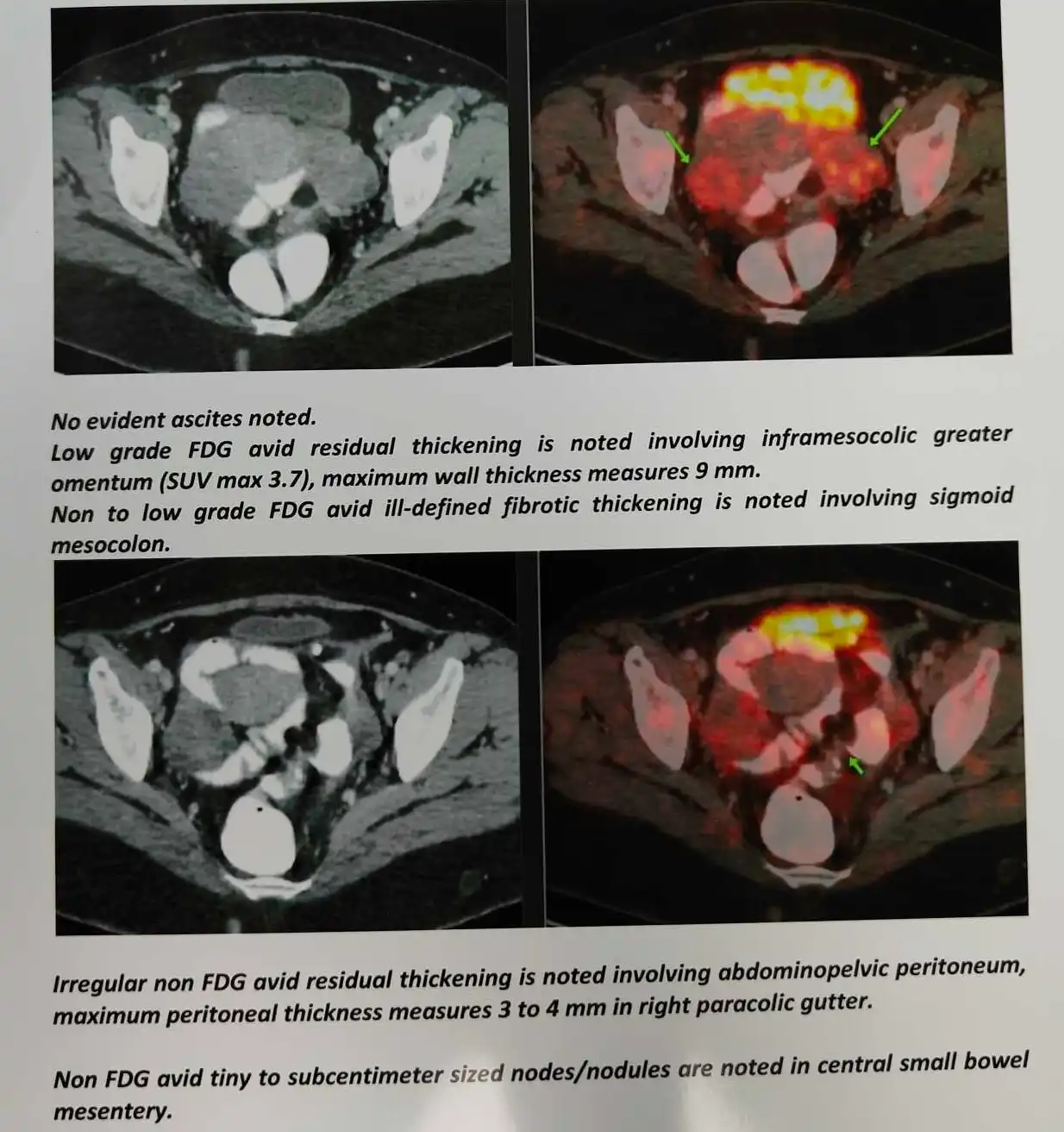
- At surgery, the importance of these residual deposits became clear. Despite their “burnt-out” appearance after chemotherapy, deposits remained over:
⦿ Both ovaries
⦿ Uterine surface
⦿ Cul-de-sac and rectosigmoid region
⦿ Omentum
⦿ Small-bowel/ileocaecal surfaces
⦿ Mesentery
⦿ Hepatoduodenal ligament
⦿ Upper peritoneum
⦿ The operation removed these visible sites of disease through extensive cytoreduction followed by HIPEC.
- Why surgery after chemotherapy?
Chemotherapy can dramatically reduce tumour volume and metabolic activity—but residual peritoneal deposits may remain.
If systemic therapy is stopped while viable peritoneal disease remains, those deposits can become the source of subsequent progression.
- Therefore, in appropriately selected patients:
Chemotherapy → reassessment of resectability → cytoreductive surgery → HIPEC
can provide a strategy for achieving the lowest possible residual tumour burden.
The objective of cytoreductive surgery is complete macroscopic cytoreduction (CC-0) whenever safely achievable. In this patient, extensive surgery reduced the documented PCI from 26 to 2, although the operative record classified the cytoreduction as CC-1.
Indian Ovarian Cancer Institute (IOCI), Ahmedabad
Every patient is unique. Treatment decisions are based on disease distribution, response to chemotherapy, resectability and multidisciplinary assessment.
Good PET Response Does Not Always Mean Good Pathological Response
In advanced ovarian cancer, radiological response and pathological response are not always the same.
This 51-year-old woman had Stage IV high-grade serous ovarian carcinoma. After 3 cycles of chemotherapy, her CA-125 fell from 4282 to 157, ascites resolved, and PET-CT showed significant regression of the ovarian, peritoneal, mesenteric and omental disease. The thoracic nodal disease showed complete metabolic resolution.
The imaging therefore suggested a good response to chemotherapy.
However, the final surgical pathology told a different story.
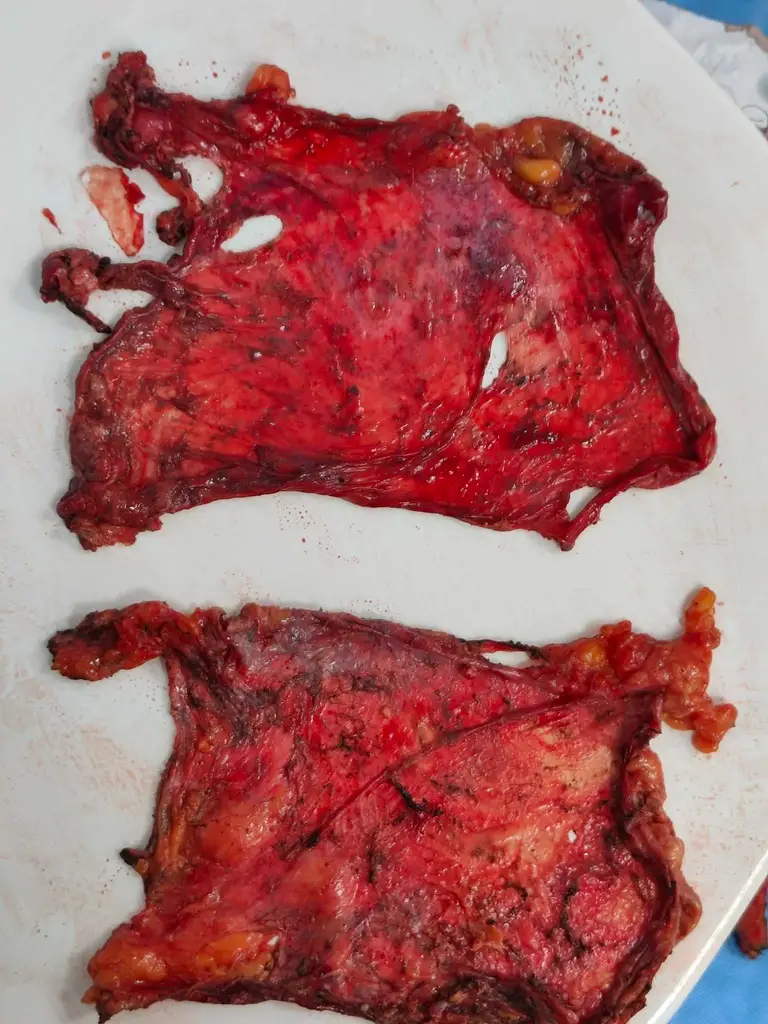
Despite the extensive “burnt-out” appearance of disease at surgery, histopathology showed Chemotherapy Response Score (CRS) 1 — minimal response. Tumour was still identified in multiple peritoneal sites, omentum, bowel/mesenteric deposits and regional lymph nodes.
- In fact, 12 of 26 examined lymph nodes contained metastatic tumour :
⦿ Right pelvic: 4/6 positive
⦿ Left pelvic: 4/5 positive
⦿ Retroperitoneal: 4/15 positive
- The learning point
A falling CA-125 and improving PET-CT are encouraging—but they do not necessarily mean that all viable cancer has disappeared.
Imaging assesses size and metabolic activity.
Pathology assesses actual tumour response at the cellular level.
This is why, in selected patients with advanced ovarian cancer, surgery remains important even after an apparently excellent response to chemotherapy.
The final treatment strategy should integrate:
Clinical response + CA-125 + imaging + laparoscopic/operative assessment + pathology
rather than relying on any single parameter.
Indian Ovarian Cancer Institute (IOCI), Ahmedabad
In ovarian cancer, response assessment is multidimensional—and treatment decisions should be individualized.
Omental Caking: What Does It Mean in Ovarian Cancer?
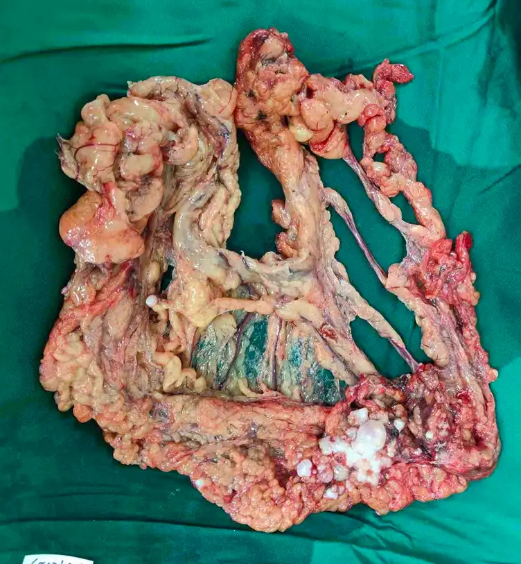
A recently managed case at IOCI highlights an important imaging feature of advanced ovarian cancer—omental caking. Ovarian cancer commonly spreads across the peritoneal surfaces. Multiple tumour deposits can infiltrate and replace the normal fatty omentum, producing nodular omental thickening or confluent “omental cake.”
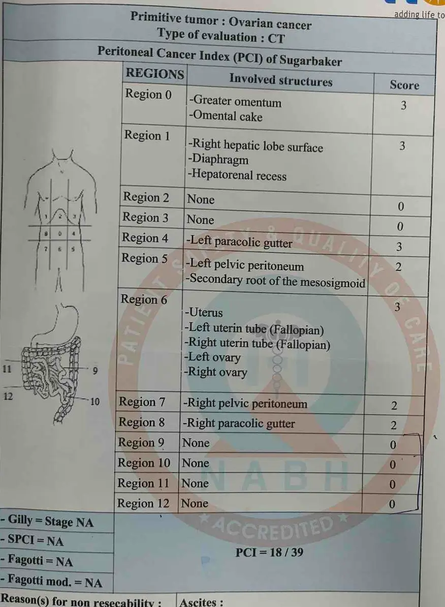
In this patient, CT showed bilateral complex solid-cystic adnexal masses, pelvic peritoneal nodules, nodular infracolic omental thickening, deposits along bilateral paracolic gutters, right subdiaphragmatic deposits and liver capsular deposits. CT PCI was 18/39.
However, imaging cannot establish the final diagnosis or completely determine resectability. At staging laparoscopy, the operative PCI was 27/39, with additional small deposits in the mesentery and mesoappendix.
The omental nodule was sent for frozen section, which confirmed metastatic adenocarcinoma and allowed definitive cytoreductive surgery to proceed.
Key learning:
Omental caking strongly suggests peritoneal spread, but it does not automatically mean inoperable disease. Imaging defines the extent; surgical assessment determines resectability, and histopathology/frozen section establishes diagnosis when required.
Stage III Ovarian Cancer: Surgery First or Chemotherapy First?
⦿ A recently managed IOCI case demonstrates why stage alone should not decide the sequence of treatment. The patient had bilateral ovarian masses, pelvic and omental deposits, bilateral subdiaphragmatic disease, pelvic nodes and retroperitoneal nodes. Yet the small-bowel serosa was free, with no gross involvement of stomach, liver or spleen.
⦿ The crucial question was: Can complete cytoreduction be safely achieved?
Staging laparoscopy was performed before committing to major surgery. Operability was confirmed, and the patient underwent extensive cytoreductive surgery (CRS)—pelvic and upper abdominal peritonectomy, bilateral pelvic lymph-node dissection, RPLND, omentectomy, appendicectomy and omphalectomy—followed by HIPEC.
⦿ The preoperative PCI was 27/39, yet postoperative PCI was 0, demonstrating complete macroscopic cytoreduction.
Key learning:
Advanced or stage III ovarian cancer does not automatically require neoadjuvant chemotherapy. When the patient is fit and complete cytoreduction appears achievable, primary surgery can be an appropriate strategy. The decision should be based on disease distribution, resectability and patient fitness—not stage or tumour size alone.
Bilateral Ovarian Masses: Always Think Beyond the Ovary
⦿ Bilateral ovarian masses require careful evaluation because they may represent either primary tubo-ovarian cancer or metastatic disease to the ovaries, particularly from the gastrointestinal tract.
⦿ In a recently managed IOCI case, CT demonstrated bilateral complex solid-cystic adnexal lesions, pelvic peritoneal deposits, omental thickening and upper abdominal peritoneal disease.
⦿ When bilateral ovarian tumours are encountered, the possibility of a gastrointestinal primary should be considered, particularly when imaging, symptoms, tumour markers or pathology raise suspicion. In such selected cases, upper GI endoscopy and colonoscopy may be required before definitive surgery to exclude a gastrointestinal primary.
⦿ In this patient, comprehensive surgical pathology ultimately demonstrated high-grade serous carcinoma involving both ovaries, with ovarian surface involvement, pelvic and omental implants. The fallopian tubes showed STIC (serous tubal intraepithelial carcinoma).
⦿ Importantly, 57 lymph nodes were examined and all were negative for metastasis, while peritoneal and omental disease accounted for the advanced stage.
Key learning:
Bilateral ovarian masses should not be labelled as primary ovarian cancer without appropriate evaluation. Imaging + tumour markers + pathology/IHC + GI evaluation when indicated help establish the origin and guide the correct surgical and oncological treatment.
PCI 27/39: Does a High PCI Mean Ovarian Cancer Is Inoperable?
A recently managed case at the Indian Ovarian Cancer Institute (IOCI), Ahmedabad underwent Staging Diagnostic Laparoscopy before definitive surgery.
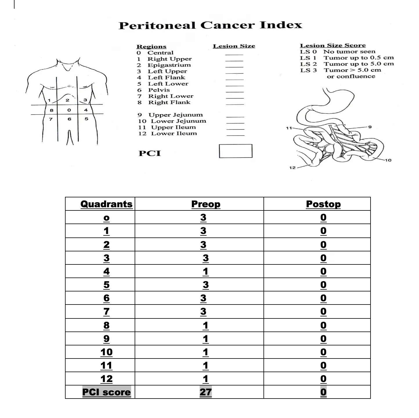
⦿ The Peritoneal Cancer Index (PCI) maps the distribution and size of peritoneal tumour deposits across 13 abdominal regions. The maximum score is 39.
⦿ In this recently managed IOCI case, CT estimated the PCI at 18/39. At staging laparoscopy, the actual operative PCI was 27/39, reflecting extensive disease involving the pelvis, omentum, bilateral subdiaphragmatic regions, paracolic areas and mesenteric sites.
⦿ But here is the important ovarian cancer message: there is no single PCI cutoff that automatically defines unresectability. PCI describes disease burden; location and resectability matter more than the number alone. Extensive disease at critical sites—such as diffuse small-bowel/mesenteric involvement or major unresectable vascular involvement—may prevent complete cytoreduction.
⦿ In this patient, despite PCI 27, complete cytoreduction was achievable. Extensive CRS was performed and the postoperative PCI was 0.
⦿ Pathology confirmed high-grade serous carcinoma, with the largest extrapelvic peritoneal deposit measuring 5.5 × 4 cm. 57/57 lymph nodes were negative, and final classification was ypT3cN0.
Key learning: PCI is a map, not a surgery “yes/no” score. In ovarian cancer, the real goal is complete cytoreduction (CC-0) whenever safely achievable.
Stage IV Ovarian Cancer: When Chemotherapy Can Make Surgery Possible
⦿ Stage IV ovarian cancer does not always mean that surgery is impossible.
⦿ In this recently managed case at IOCI, the patient had extensive ovarian cancer with bilateral adnexal disease, peritoneal/omental involvement, pelvic and retroperitoneal lymph nodes, ascites and pleural effusion.
⦿ The initial disease burden made neoadjuvant chemotherapy (NACT) the appropriate strategy. After 3 cycles of Paclitaxel + Carboplatin, with Bevacizumab during the first 2 cycles, there was a remarkable response.
⦿ CA-125 fell from 1978 to 43.7 U/mL. Imaging showed reduction in bilateral adnexal lesions, pelvic and retroperitoneal lymph nodes, omental/mesenteric disease and ascites, with resolution of the previously noted pleural effusion.
⦿ The patient was subsequently taken for interval cytoreductive surgery, where extensive disease could be systematically addressed.
Key learning:
NACT is not simply postponing surgery. In appropriately selected advanced ovarian cancer patients, a good chemotherapy response can convert extensive disease into a setting where complete cytoreduction becomes achievable.
CC-0 Cytoreduction: The Real Goal of Advanced Ovarian Cancer Surgery
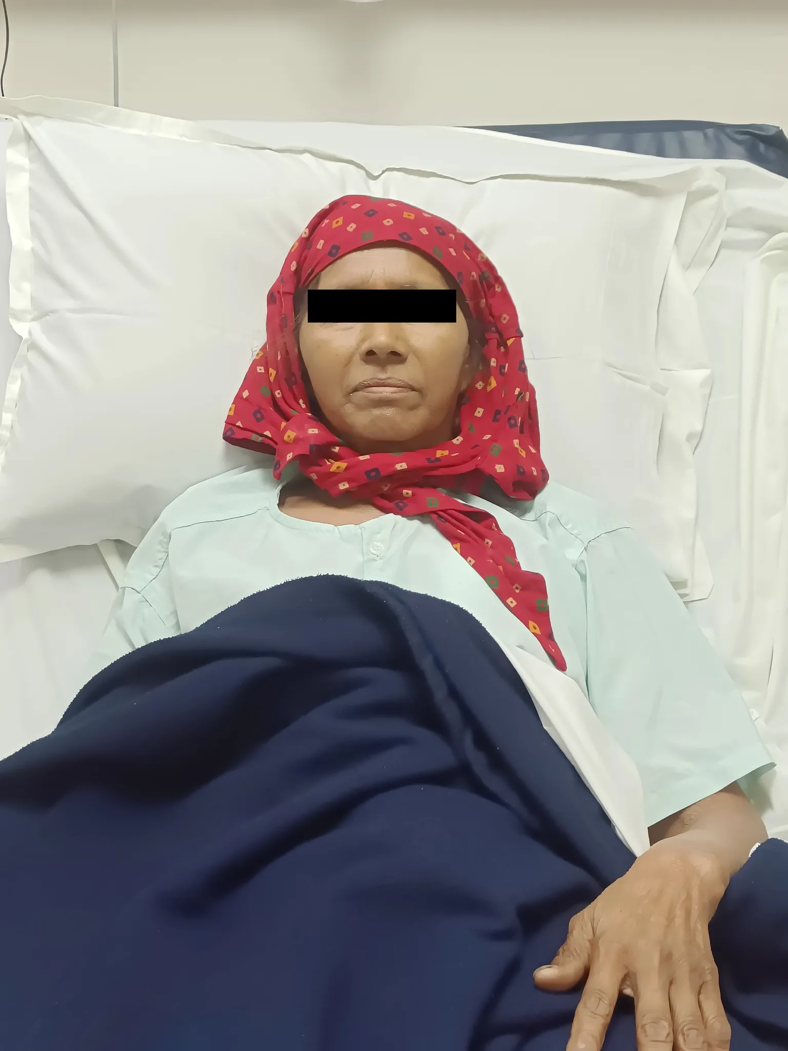
⦿ In this recently managed case at IOCI, chemotherapy produced substantial regression of the previously extensive disease. At surgery, residual disease involved the adnexa, pelvis, mesentery and omentum, along with treated/“burnt-out” upper abdominal deposits.
⦿ A comprehensive cytoreductive surgery (CRS) was performed, addressing disease across multiple compartments. The operative Peritoneal Cancer Index (PCI) was 9, and after cytoreduction the residual disease score was 0, indicating CC-0 complete macroscopic cytoreduction. This distinction is critical.
⦿ A patient may initially have widespread peritoneal disease, but if the disease is responsive to chemotherapy and technically resectable, complete cytoreduction can still be achieved.
Key learning:
In advanced ovarian cancer, the important surgical endpoint is no visible residual disease, because incomplete cytoreduction can compromise the benefit of aggressive treatment.
Ovarian Tumour Adherent to Sigmoid: Adhesion Does Not Always Mean Bowel Resection
Does an ovarian cancer attached to the sigmoid colon automatically require bowel removal? Not always.
In this recently managed case at IOCI, the ovarian/peritoneal disease was closely related to the sigmoid region, with involvement around the rectosigmoid/sigmoid mesentery. Imaging had already demonstrated loss of normal fat planes and close bowel proximity.
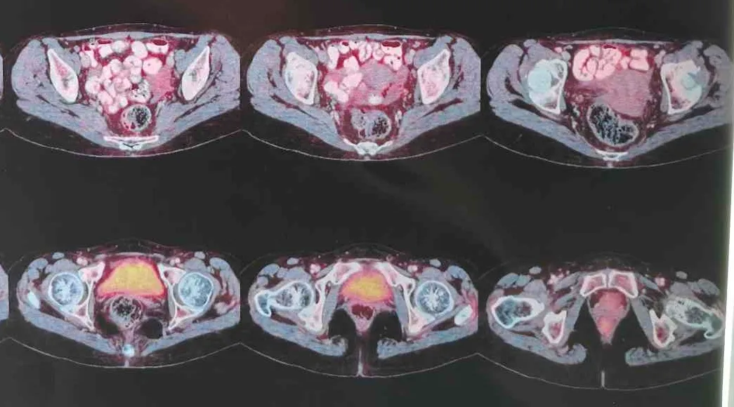
At surgery, meticulous pelvic cytoreduction was performed. The surgical objective was to remove all visible cancer while preserving uninvolved bowel whenever an oncologically safe plane could be developed.
This highlights an important principle in advanced ovarian cancer surgery:
“Adherent” does not always mean “invading.”
Cancer may be attached to bowel because of inflammation, fibrosis, tumour deposits or true bowel-wall invasion. The surgeon must carefully assess the plane and determine whether bowel resection is genuinely necessary.
With meticulous dissection, sharp adhesiolysis and careful assessment of bowel involvement, bowel preservation may be possible in selected patients.
Key learning:
The goal of CRS is complete cancer clearance with maximum preservation of organ function whenever oncologically safe. Expert pelvic oncology surgery can sometimes avoid unnecessary bowel resection—even when ovarian cancer appears densely adherent to the sigmoid.
What Happens After Neoadjuvant Chemotherapy? Pathology Gives the Final Answer
⦿ Imaging shows response. Surgery shows resectability. Histopathology tells us what cancer remains.
⦿ In this recently managed case at IOCI, the final pathology confirmed high-grade serous carcinoma involving both ovaries, with residual microscopic peritoneal/omental disease after chemotherapy.
⦿ The specimen demonstrated a Chemotherapy Response Score (CRS) 2, indicating a moderate response to neoadjuvant chemotherapy. Importantly, 54 lymph nodes were examined and all were negative (0/54). The final pathological classification was ypT3aN0.
⦿ This illustrates why several parameters must be considered together after interval cytoreductive surgery: Clinical response + radiological response + completeness of cytoreduction + chemotherapy response score + nodal status + final pathological stage.
⦿ No single parameter tells the entire story.
⦿ The patient had initially extensive disease, but after chemotherapy and comprehensive cytoreduction, there was no gross residual disease, with negative regional lymph nodes.
Key learning:
Pathology after NACT is not merely a diagnosis—it provides important information about tumour biology, chemotherapy response, residual disease and prognosis, helping guide subsequent ovarian cancer management.
Get health updates from Dr Swati Shah on WhatsApp
Swasth Parivar: twice a month, practical advice on recovery, diet and warning signs. No advertising. Stop any time.
Your number is used for these updates and for the clinic to reach you. It is never sold or passed on. To stop, reply STOP on WhatsApp.