


Case Library - Recurrent Ovary Cancer
Endometrioid Ovarian Cancer with Suspected Residual Pelvic Disease After Previous Surgery
Endometrioid carcinoma is a less common subtype of epithelial ovarian cancer. In some patients, further evaluation after the initial surgery may identify findings that require additional treatment. In one case managed at the Indian Ovarian Cancer Institute (IOCI), Ahmedabad, the patient had previously undergone total abdominal hysterectomy with bilateral salpingo-oophorectomy for endometrioid ovarian carcinoma. Subsequent PET-CT and operative assessment suggested pelvic peritoneal deposits, scar nodules and an omental nodule, leading to further surgical evaluation.
The patient underwent comprehensive cytoreductive surgery, including pelvic peritonectomy, cul-de-sactomy, omentectomy, pelvic and para-aortic lymph node dissection, upper peritoneal biopsies, revision of the previous surgical scar and removal of suspicious nodules. Histopathological examination subsequently demonstrated postoperative inflammatory and healing-related changes without viable tumour cells in the revised scar, pelvic peritoneum, omentum or upper peritoneal biopsies. All examined pelvic and para-aortic lymph nodes were negative for metastasis.
Clinical Insight:
Imaging or operative findings that appear suspicious after previous ovarian cancer surgery do not always represent active cancer. Definitive diagnosis requires histopathological examination of the removed tissues.
Why Cytoreductive Surgery Was Planned After PET-CT Findings
Treatment planning after ovarian cancer surgery often depends on new imaging findings. In this case, PET-CT demonstrated two metabolically active lesions—one adjacent to the rectosigmoid region and another in the suprapubic region. The patient also had a recent diagnosis of endometrioid ovarian carcinoma following previous surgery, prompting multidisciplinary evaluation for possible residual disease.
Based on these findings, cytoreductive surgery was planned to evaluate and remove suspected disease. During surgery, suspicious areas involving the pelvic peritoneum, previous surgical scar, infundibulopelvic ligament region, vault and omentum were excised. Histopathology later showed that these suspicious areas represented chronic inflammation, foreign body giant cell reaction, suture granuloma and postoperative tissue changes, with no evidence of viable tumour cells.
Clinical Insight:
PET-CT is an important tool for identifying suspicious lesions, but imaging alone cannot confirm active cancer. Surgical assessment and histopathological examination remain the gold standard for distinguishing residual malignancy from postoperative inflammatory changes.
This clinical experience is shared by Indian Ovarian Cancer Institute (IOCI), Ahmedabad.
Every patient is unique. Treatment recommendations are individualized based on clinical evaluation, imaging, pathology and multidisciplinary discussion.
Cytoreductive Surgery for Suspected Residual Endometrioid Ovarian Cancer
Managing suspected residual ovarian cancer may require meticulous exploration of multiple abdominal and pelvic regions. In this case, cytoreductive surgery included exploratory laparotomy, pelvic peritonectomy, cul-de-sactomy, revision of the infundibulopelvic ligament and vault, greater omentectomy, bilateral pelvic lymph node dissection, para-aortic lymph node dissection, upper peritoneal biopsies, umbilectomy and revision of the previous surgical scar.
During surgery, deposits were noted around the previous scar, pelvic peritoneum, infundibulopelvic ligament region and omentum, while several upper abdominal organs appeared grossly normal. Comprehensive excision allowed detailed pathological assessment of all suspicious areas. Histopathology ultimately showed no viable tumour in the excised scar, omentum, pelvic peritoneum or upper peritoneal biopsies, and all 65 examined pelvic and para-aortic lymph nodes were free of metastasis.
Clinical Insight:
Cytoreductive surgery is valuable not only for tumour removal but also for accurately determining whether suspicious postoperative findings represent persistent cancer or benign healing-related changes.
This educational case is shared by Indian Ovarian Cancer Institute (IOCI), Ahmedabad.
Every patient is unique. Treatment recommendations are individualized based on clinical evaluation, imaging, pathology and multidisciplinary discussion.
When Histopathology Shows Healing Rather Than Residual Cancer
Histopathology plays a critical role after surgery for ovarian cancer, especially when imaging or operative findings raise concern for persistent disease. In this case, multiple tissues that appeared suspicious during surgery—including the revised scar, pelvic peritoneum, omentum and upper peritoneal biopsy—were carefully examined under the microscope.
Rather than demonstrating active malignancy, the pathology report described chronic inflammation, foreign body giant cell reaction, suture granuloma, fat necrosis and calcification, findings that are consistent with postoperative healing changes. No viable tumour cells were identified in these specimens. In addition, twenty left pelvic lymph nodes, fifteen right pelvic lymph nodes and thirty para-aortic lymph nodes all showed reactive features without metastatic disease.
Clinical Insight:
Postoperative inflammation, scar tissue and suture-related reactions can sometimes resemble recurrent disease on imaging or during surgery. Histopathological examination is essential to distinguish true recurrence from benign postoperative changes and to guide future management.
This case highlights the importance of comprehensive pathological assessment at Indian Ovarian Cancer Institute (IOCI), Ahmedabad.
Every patient is unique. Treatment recommendations are individualized based on clinical evaluation, imaging, pathology and multidisciplinary discussion.
Can Recurrent Ovarian Cancer Still Be Curable? Selected Patients May Benefit from Complete Secondary Cytoreduction
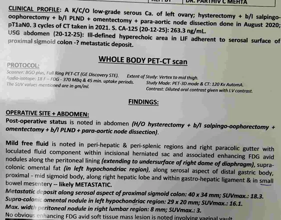
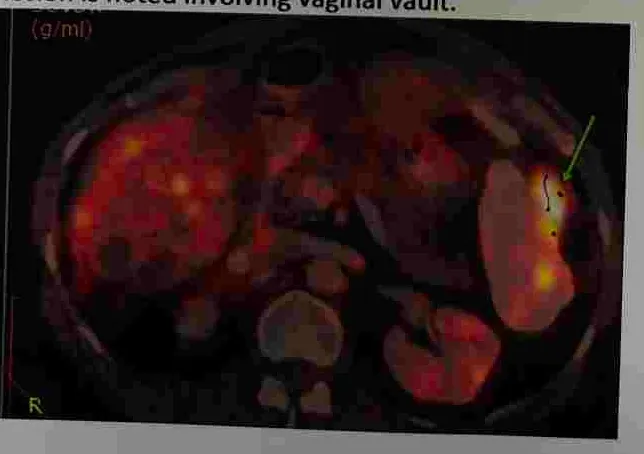
Five years after initial treatment for ovarian cancer, this patient developed recurrent disease. PET-CT demonstrated metabolically active deposits along the peritoneal lining extending to the undersurface of the right hemidiaphragm, supracolic omentum, distal stomach, proximal sigmoid colon, gastrohepatic ligament, right hepatic surface and small bowel mesentery, together with mild malignant pleural disease.
Following three cycles of paclitaxel-carboplatin chemotherapy, repeat PET-CT showed complete resolution of ascites, complete resolution of metastatic pleural thickening, no fresh hypermetabolic lesions, and near-static metabolic activity of the remaining peritoneal deposits without new distant systemic metastases. Although a CT comparison described mild increase in the size of a sigmoid deposit and left lumbar omental disease, there was simultaneous regression of ascites and mesenteric haziness, indicating a mixed radiological response rather than uncontrolled dissemination.
Because disease remained confined to surgically resectable peritoneal sites, secondary cytoreductive surgery followed by HIPEC was performed.
Clinical Insight
Recurrent ovarian cancer is not always incurable. In carefully selected patients with localized, resectable recurrence and good response to systemic therapy, complete secondary cytoreduction may provide meaningful long-term disease control and, in selected situations, prolonged survival.
Indian Ovarian Cancer Institute (IOCI), Ahmedabad
Every patient is unique. Treatment recommendations are individualized based on clinical evaluation, imaging, pathology and multidisciplinary discussion.
When CT Suggests Progression After Chemotherapy, Has the Treatment Really Failed?
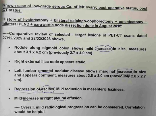
After three cycles of chemotherapy, CT comparison reported a mild increase in the sigmoid deposit (from 2.7 × 4.0 cm to 3.1 × 4.2 cm) and enlargement of left lumbar omental disease, leading to an overall impression of “mild radiological progression.” At the same time, however, ascites had regressed and mesenteric haziness had improved. PET-CT also demonstrated complete resolution of metastatic pleural thickening, complete resolution of ascites, and no new hypermetabolic metastatic lesions.
During surgery, these apparently enlarging lesions were repeatedly described as “burnt-out deposits”—a 4 × 4 cm burnt-out intramural sigmoid mass, burnt-out deposits over the rectosigmoid serosa and mesentery, and multiple tiny burnt-out deposits over bowel surfaces and the upper peritoneum.
Chemotherapy can destroy viable tumour cells, leaving behind fibrosis, necrosis or acellular tumour beds that may not shrink immediately on imaging. Therefore, treatment response should never be judged solely by lesion size. Decisions regarding further chemotherapy versus surgery should integrate metabolic imaging, tumour markers, disease distribution and operability—not RECIST measurements alone.
Clinical Insight
The objective of neoadjuvant chemotherapy is not only tumour shrinkage. It also treats microscopic systemic disease while converting viable peritoneal deposits into surgically removable residual disease.
Indian Ovarian Cancer Institute (IOCI), Ahmedabad
Every patient is unique. Treatment recommendations are individualized based on clinical evaluation, imaging, pathology and multidisciplinary discussion.
Secondary Cytoreductive Surgery with HIPEC for Recurrent Ovarian Cancer
At surgery, the Peritoneal Cancer Index (PCI) was 26, demonstrating extensive recurrent peritoneal disease. Disease involved the supracolic omentum, sigmoid colon, rectosigmoid mesentery, cul-de-sac, right upper peritoneum, left upper peritoneum, hernial sac and multiple small bowel mesenteric deposits. Operability was confirmed before proceeding with complete cytoreductive surgery.
The operation included pelvic peritonectomy, bilateral upper peritonectomy, segmental sigmoid colectomy, supracolic omentectomy, lesser omentectomy, falciform ligament excision, excision of bowel surface deposits, mesenteric deposits and hernial sac deposits, followed by HIPEC using cisplatin 150 mg at 43°C for 90 minutes. Post-cytoreduction PCI was reduced to CC1, indicating only minimal residual disease.
Histopathology confirmed residual high-grade serous carcinoma involving the omentum, sigmoid colon, appendix, upper peritoneum, hernial sac and mesenteric deposits, while both bowel resection margins remained free of tumour.
Clinical Insight
Secondary CRS with HIPEC is not performed simply because recurrence is present. The goal is complete or near-complete cytoreduction in carefully selected patients whose recurrent disease remains surgically resectable.
Indian Ovarian Cancer Institute (IOCI), Ahmedabad
Every patient is unique. Treatment recommendations are individualized based on clinical evaluation, imaging, pathology and multidisciplinary discussion.
Does Bowel Involvement in Ovarian Cancer Always Mean a Permanent Stoma?
PET-CT demonstrated recurrent disease along the serosal aspect of the proximal-mid sigmoid colon, while surgery confirmed an approximately 4 × 4 cm intramural burnt-out sigmoid mass with multiple deposits over the rectal and sigmoid serosa and mesentery.
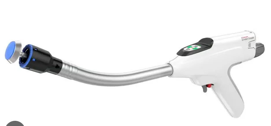
Because the tumour extended into the bowel wall, segmental sigmoid colectomy was required. Instead of creating a permanent stoma, continuity of the intestine was restored immediately using an end-to-end stapled colo-colic anastomosis. Additional tumour deposits were meticulously removed from the transverse colon, descending colon, small bowel surface and mesentery as part of complete cytoreduction.
Histopathology confirmed metastatic high-grade serous carcinoma within the sigmoid segment, while both the proximal and distal bowel margins and the anastomotic doughnuts were free of tumour.
Clinical Insight
Ovarian cancer frequently spreads to the surface of the rectum and sigmoid colon because it is a peritoneal disease. Bowel resection may therefore be necessary during cytoreductive surgery. However, with meticulous technique, appropriate blood supply and tension-free stapled anastomosis, many patients can avoid a permanent stoma.
Indian Ovarian Cancer Institute (IOCI), Ahmedabad
Every patient is unique. Treatment recommendations are individualized based on clinical evaluation, imaging, pathology and multidisciplinary discussion.
Recurrent Ovarian Cancer — When Can Surgery Still Aim for Complete Disease Clearance?
A recently managed case at the Indian Ovarian Cancer Institute (IOCI), Ahmedabad) illustrates why recurrence of ovarian cancer does not automatically mean that treatment is limited to chemotherapy.
A 58-year-old woman with previously treated high-grade serous carcinoma of the ovary developed recurrence. Her CA-125 was 366 U/mL at recurrence. She received 3 cycles of paclitaxel + carboplatin chemotherapy, following which CA-125 fell dramatically to 21.4 U/mL. Imaging also showed regression of the retroperitoneal disease and resolution of the bowel obstruction.
With a good response to chemotherapy and potentially surgically addressable residual disease, she was evaluated for secondary cytoreductive surgery.
At surgery, the peritoneal surfaces were free of gross disease and there was no ascites. However, enlarged paracaval, retrocaval and left para-aortic lymph nodes were identified, along with a small residual mesenteric nodule. These areas were surgically addressed.
Histopathology demonstrated metastatic high-grade serous carcinoma in the retroperitoneal lymph nodes and peritoneal biopsy. 4 of 13 retroperitoneal lymph nodes were positive, with the largest metastatic deposit measuring 1.8 × 1 cm.
The important message:
Recurrent ovarian cancer is not synonymous with incurable disease.
In selected patients, a strong response to chemotherapy can create an opportunity for secondary cytoreductive surgery, particularly when the remaining disease can be completely removed.
The decision should be based on the pattern and volume of recurrence, response to systemic treatment, patient’s fitness and—most importantly—the possibility of achieving complete cytoreduction.
At IOCI, recurrent ovarian cancer is evaluated with the objective of combining systemic treatment and appropriately selected surgery to achieve the best possible long-term disease control.
Retroperitoneal Lymph Node Dissection Should Not Be Missed in Ovarian Cancer Surgery
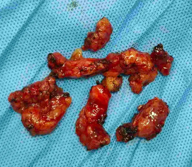
Ovarian cancer does not spread only to the pelvic lymph nodes. The ovaries have important lymphatic drainage to the para-aortic and retroperitoneal lymphatic chains. Therefore, comprehensive ovarian cancer surgery must always address the retroperitoneal lymphatic compartment and should not focus only on the pelvis.
A recently managed recurrent ovarian cancer case at IOCI, Ahmedabad, demonstrates this clearly. After chemotherapy, imaging showed regression of the retroperitoneal lymph nodes. During secondary cytoreductive surgery, however, enlarged paracaval and retrocaval nodes and left para-aortic lymph nodes were identified. A retroperitoneal lymph node dissection (RPLND) was performed.
- The pathology proved the importance of removing and examining these nodes:
4 out of 13 retroperitoneal lymph nodes contained metastatic high-grade serous carcinoma, with the largest metastatic deposit measuring 1.8 × 1 cm.
This means that even when the peritoneal surfaces appear clear, clinically important ovarian cancer can remain within the retroperitoneal lymphatic system.
- Why does this matter?
• Pelvic disease alone does not represent the complete lymphatic spread pattern of ovarian cancer.
• Para-aortic/retroperitoneal nodes can harbour metastatic disease even when other peritoneal surfaces are clear.
• Removing suspicious or involved retroperitoneal nodes provides definitive pathological information.
• RPLND is technically demanding and requires experience with major vessels, ureters and retroperitoneal structures.
The surgical principle:
In ovarian cancer surgery, retroperitoneal lymph-node assessment should always be addressed. It should not be omitted simply because the visible disease is predominantly pelvic or peritoneal.
At IOCI, comprehensive ovarian cancer surgery includes systematic assessment of pelvic and retroperitoneal disease, with RPLND performed when indicated and technically appropriate.
Second Ovarian Cancer Surgery Does Not Mean Poor Quality of Life
- One of the biggest concerns patients have after ovarian cancer recurrence is:
“If I need another major surgery, how will I recover?”
A recently managed patient at IOCI, Ahmedabad, underwent secondary cytoreductive surgery for recurrent high-grade serous ovarian cancer after responding well to chemotherapy.
The procedure included exploratory laparotomy, adhesiolysis, retroperitoneal lymph node dissection and peritoneal/mesenteric biopsies. Multiple bowel loops were adherent to each other and to the previous midline scar, making adhesiolysis an important part of the procedure.
Despite this being a second major abdominal cancer operation, the postoperative course was satisfactory.
There was a transient period of low urine output, which was managed conservatively with intravenous fluids and albumin. The urinary catheter was subsequently removed, the drain was removed, and the patient was discharged in a stable, ambulatory condition with a clean wound and controlled pain.
The operation itself had a recorded blood loss of only 100 mL, with no blood transfusion documented in the operation note.
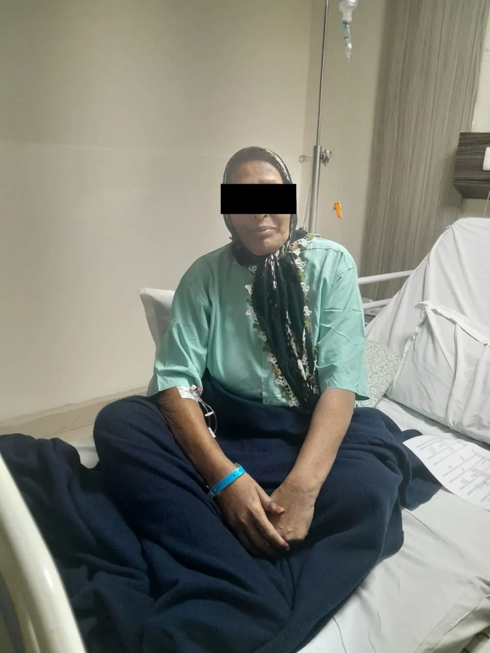
The message for patients:
Do not assume that a second ovarian cancer surgery necessarily means prolonged dependence or poor quality of life.
With careful patient selection, meticulous surgical technique, appropriate perioperative care and an experienced multidisciplinary team, even complex secondary cytoreductive procedures can be followed by satisfactory recovery.
Cancer treatment should not be judged only by whether an operation was possible.
Recovery, independence, return to activity and quality of life are equally important goals.
At IOCI, surgical planning for recurrent ovarian cancer therefore considers both oncological benefit and functional recovery.
Get health updates from Dr Swati Shah on WhatsApp
Swasth Parivar: twice a month, practical advice on recovery, diet and warning signs. No advertising. Stop any time.
Your number is used for these updates and for the clinic to reach you. It is never sold or passed on. To stop, reply STOP on WhatsApp.