


Case Library - 1st stage Ovary cancer
When a Complex Ovarian Mass on MRI Raises Concern for Ovarian Cancer
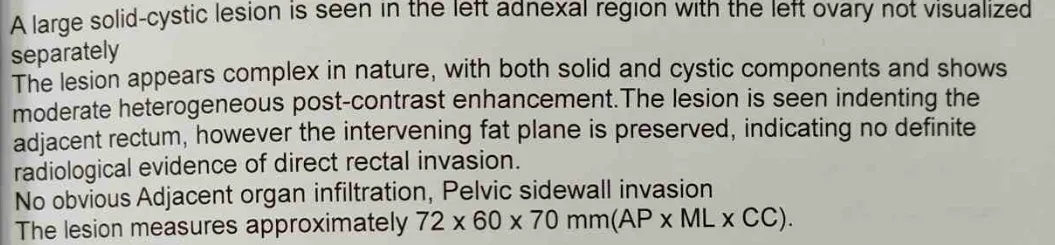
A woman was evaluated for a complex left ovarian mass after imaging identified a multiloculated solid-cystic lesion with enhancing solid components. MRI classified the lesion as O-RADS MRI 5, indicating a high likelihood of malignancy. The lesion measured approximately 7.7 × 6.0 × 6.2 cm, while the right ovary appeared normal and there was no significant pelvic lymph node enlargement on imaging.
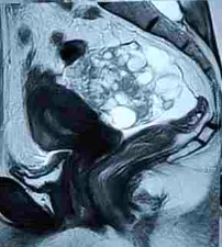
⦿ Following multidisciplinary evaluation, surgical management was planned. Staging laparoscopy was initially performed, followed by conversion to open surgery after identifying an ovarian mass with surface deposits and small pelvic peritoneal deposits. The patient underwent complete surgical staging including removal of the ovarian tumour, hysterectomy, bilateral pelvic and retroperitoneal lymph node dissection, omentectomy and peritoneal biopsies.
- Clinical Insight
A complex ovarian mass containing both cystic and solid components requires careful evaluation. MRI helps estimate the risk of malignancy, but the final diagnosis depends on surgery and histopathological examination.
This educational case is shared by Indian Ovarian Cancer Institute (IOCI), Ahmedabad.
Every patient is unique. Treatment recommendations are individualized based on clinical evaluation, imaging, pathology and multidisciplinary discussion.
Why Staging Laparoscopy Was Performed Before Definitive Ovarian Cancer Surgery
⦿ Choosing the most appropriate surgical approach is an important step in ovarian cancer management. In this case, imaging suggested a localized left ovarian tumour without obvious distant spread. To assess the abdominal cavity directly, staging laparoscopy was performed before proceeding with definitive surgery.
⦿ During laparoscopy, surgeons identified an approximately 8 cm left ovarian mass with surface involvement, a small amount of pelvic fluid and tiny pelvic peritoneal deposits. Based on these findings, the surgical team converted to an open procedure to perform comprehensive staging surgery. Frozen section examination of the ovarian mass confirmed invasive malignancy during the operation, allowing complete staging to be performed in the same sitting.
- Clinical Insight
Staging laparoscopy provides valuable real-time information about disease extent. In selected patients, it helps surgeons choose the safest and most appropriate operative strategy instead of relying only on imaging findings.
This clinical experience is shared by Indian Ovarian Cancer Institute (IOCI), Ahmedabad.
Every patient is unique. Treatment recommendations are individualized based on clinical evaluation, imaging, pathology and multidisciplinary discussion.
Why Removing an Ovarian Tumour Intact Is Important
During ovarian cancer surgery, preserving the integrity of the tumour whenever feasible is an important surgical principle. In this case, the left ovarian mass was carefully dissected and removed intact, together with the fallopian tube, before being sent for frozen section examination. The frozen section confirmed invasive malignancy, following which comprehensive surgical staging was completed during the same operation.
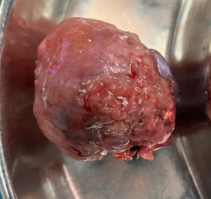
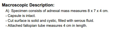
⦿ The procedure included hysterectomy, removal of the opposite ovary and fallopian tube, bilateral pelvic lymph node dissection, retroperitoneal lymph node dissection, omentectomy, peritoneal biopsies and fluid cytology. Histopathology later described an 8 × 7 × 4 cm tumour with an intact capsule, allowing detailed pathological assessment.
- Clinical Insight
Removing an ovarian tumour intact helps preserve tissue architecture for accurate pathological evaluation and reduces the risk of intraoperative tumour spillage whenever technically feasible.
This educational case is shared by Indian Ovarian Cancer Institute (IOCI), Ahmedabad.
Every patient is unique. Treatment recommendations are individualized based on clinical evaluation, imaging, pathology and multidisciplinary discussion.
Why Histopathology Changed the Final Stage of This Ovarian Cancer
⦿ Imaging suggested a localized ovarian tumour, but the final stage of ovarian cancer is determined only after complete pathological examination of all tissues removed during surgery.
⦿ Histopathology confirmed an intermediate- to high-grade adenocarcinoma arising from the left ovary measuring 8 × 7 × 4 cm. The right ovary, fallopian tubes, uterus, omentum and peritoneal biopsies were free of tumour. However, examination of 31 lymph nodes revealed 2 metastatic retroperitoneal lymph nodes, while all pelvic lymph nodes were negative. Based on these findings, the tumour was classified as FIGO Stage IIIA1(i). Immunohistochemistry was also recommended for further tumour subtyping.
- Clinical Insight
The final stage of ovarian cancer depends not only on the ovarian tumour itself but also on whether cancer has spread to lymph nodes or other tissues. Comprehensive surgical staging and histopathological examination are essential for accurate staging and planning further treatment.
This educational case is shared by Indian Ovarian Cancer Institute (IOCI), Ahmedabad.
Every patient is unique. Treatment recommendations are individualized based on clinical evaluation, imaging, pathology and multidisciplinary discussion.
CA-125, AFP, β-hCG & LDH Were Normal… Why Was Inhibin Tested in This 12-Year-Old Girl with a Giant Ovarian Tumour?
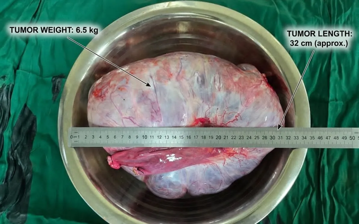
Not every ovarian tumour produces the commonly known tumour markers. A 12-year-old girl evaluated at the Indian Ovarian Cancer Institute (IOCI), Ahmedabad presented with a giant ovarian mass. Surprisingly, routine tumour markers including CA-125, AFP, β-hCG and LDH were not suggestive of the diagnosis. However, because the patient’s age, imaging findings and clinical presentation raised suspicion for a sex cord stromal tumour, Serum Inhibin was requested before surgery and was markedly elevated (>1500 pg/mL).
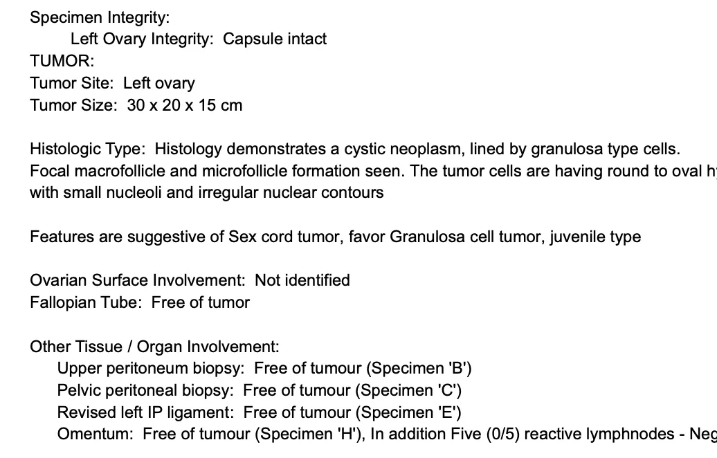
⦿ The ovarian tumour was removed intact, and frozen section suggested a malignant sex cord stromal tumour. Final histopathology confirmed a Juveline Granulosa Cell Tumour measuring 30 × 20 × 15 cm, with disease confined to the ovary (FIGO Stage IA). Forty-one lymph nodes, peritoneal biopsies and omentum were free of tumour.
- Expert Insight
When a child or young woman has a large ovarian tumour with normal CA-125, AFP, β-hCG and LDH, clinicians should also consider granulosa cell tumour among the differential diagnoses. Serum Inhibin, particularly Inhibin B, is an important tumour marker that can help support the diagnosis and is also valuable during follow-up.
CT Showed a 32 cm Ovarian Tumour Compressing Both Ureters, Major Blood Vessels and Bowel—How Can It Be Removed Safely?
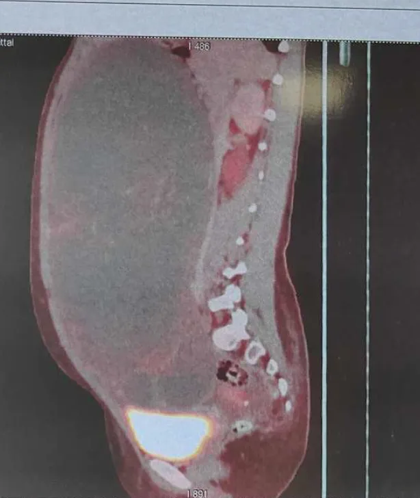
Preoperative PET-CT revealed a massive complex ovarian tumour measuring approximately 13.5 × 23.8 × 32 cm. The tumour was compressing both ureters, displacing the uterus and left ovary, compressing small bowel loops, inferior vena cava, abdominal aorta and bilateral iliac vessels, and extending from the infrahepatic region to the urinary bladder. Despite its enormous size, imaging suggested compression rather than definite invasion of these structures.
During surgery at the Indian Ovarian Cancer Institute (IOCI), Ahmedabad, meticulous retroperitoneal dissection was performed. The ureter was identified and safeguarded before ligating the infundibulopelvic ligament. The tumour was then carefully separated from adjacent organs and removed intact without rupture, while preserving the bowel, ureters and major blood vessels. Frozen section subsequently guided completion staging surgery.
Surgical Perspective
Large ovarian tumours often displace vital organs rather than infiltrate them. Understanding pelvic and retroperitoneal anatomy is essential for safe tumour removal while preserving surrounding structures whenever oncologically appropriate.
Why Is Frozen Section Called a "Live Biopsy" During Ovarian Tumour Surgery?
Patients often ask why surgeons do not simply perform a needle biopsy before operating on an ovarian mass. In many ovarian tumours, preoperative biopsy is generally avoided because puncturing the tumour may lead to rupture, alter staging and potentially influence subsequent treatment.
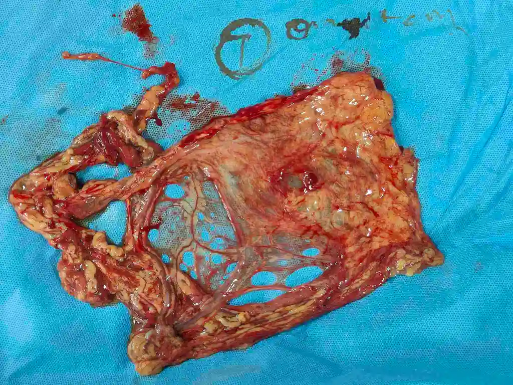
In this young patient managed at the Indian Ovarian Cancer Institute (IOCI), Ahmedabad, the ovarian tumour was first removed intact. While the patient remained under anaesthesia, the specimen was immediately examined using frozen section, a rapid pathological assessment often referred to as a “live biopsy.” Frozen section suggested a malignant sex cord stromal tumour, allowing the surgical team to proceed directly with bilateral pelvic lymph node dissection, retroperitoneal lymph node dissection, omentectomy and peritoneal biopsies during the same operation. Final histopathology later confirmed a Juveline Granulosa Cell Tumour, FIGO Stage IA.
- Did You Know?
Frozen section helps avoid unnecessary delays between diagnosis and definitive treatment. It often prevents a second operation by providing an immediate diagnosis that guides the extent of surgery.
Can Removing an Ovarian Tumour Intact Help Avoid Chemotherapy?
One of the fundamental principles of ovarian cancer surgery is to remove the tumour without rupture whenever safely possible. In this case managed at the Indian Ovarian Cancer Institute (IOCI), Ahmedabad, the ovarian tumour measured 30 × 20 × 15 cm and was removed intact with the capsule preserved. Histopathology confirmed no ovarian surface involvement, no capsular breach, negative peritoneal washings, negative omentum, negative peritoneal biopsies and 41 negative lymph nodes, resulting in FIGO Stage IA Juvenile Granulosa Cell Tumour.
- Why This Matters
International gynecologic oncology guidelines recommend avoiding tumour rupture whenever feasible because capsule rupture may upstage an otherwise Stage IA tumour to Stage IC, depending on the circumstances. This change in stage can influence postoperative treatment recommendations, including the need for adjuvant chemotherapy in selected patients.
Sometimes, choosing a larger abdominal incision is not about making surgery easier—it is about maximizing the chance of removing the tumour safely, intact and according to sound oncologic principles.
What Does an O-RADS 4 Ovarian Mass Mean? Does It Always Indicate Ovarian Cancer?
An ultrasound report mentioning O-RADS 4 often causes anxiety, but it does not automatically mean that the ovarian mass is cancer. O-RADS (Ovarian-Adnexal Reporting and Data System) is a standardized ultrasound scoring system that estimates the risk of malignancy and helps determine which patients should be evaluated by a gynecologic oncologist.
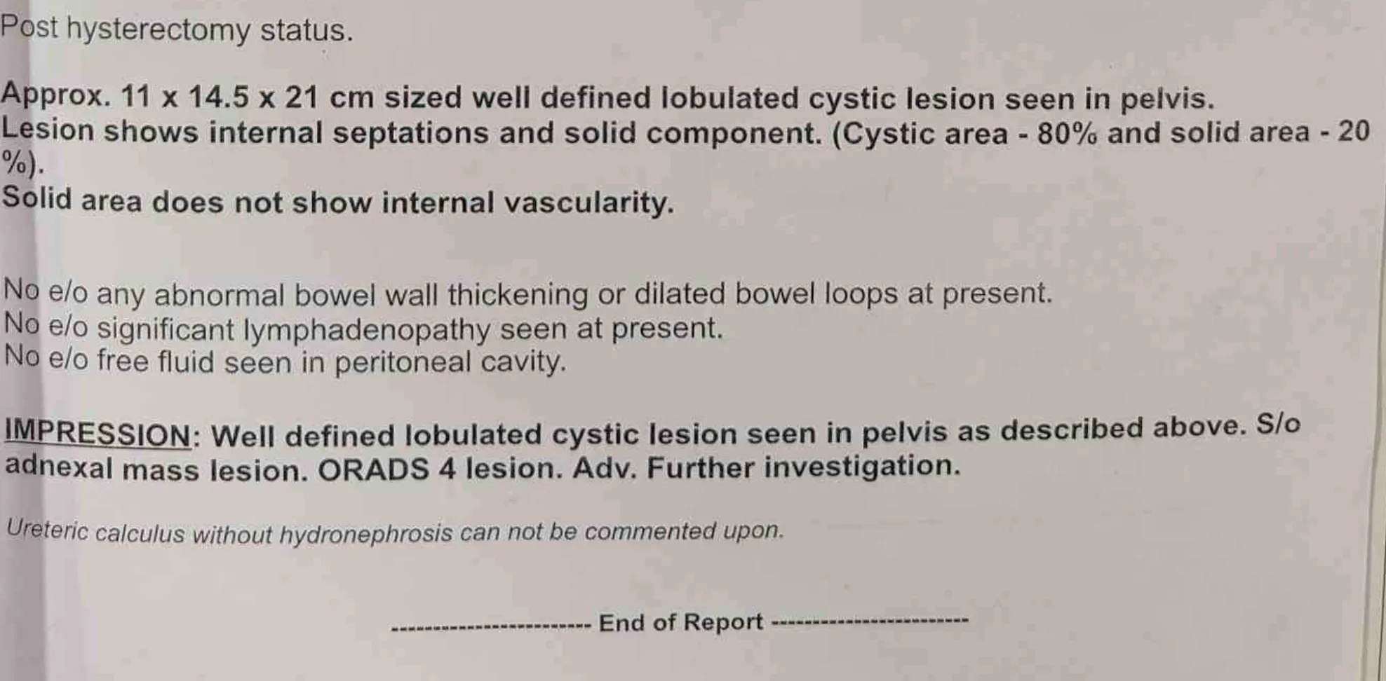
In one recently managed case at the Indian Ovarian Cancer Institute (IOCI), Ahmedabad, ultrasound demonstrated a well-defined 11 × 14.5 × 21 cm multiloculated cystic pelvic mass containing internal septations and a solid component, leading to an O-RADS 4 classification. CT scan further characterized the lesion and helped plan surgery. Final histopathology confirmed high-grade serous carcinoma of ovarian origin. This case illustrates why an O-RADS 4 lesion requires timely specialist assessment rather than observation alone.
- Clinical Insight
O-RADS is a risk assessment tool, not a final diagnosis. It helps identify ovarian masses that need further evaluation with tumour markers, advanced imaging and expert surgical planning. Early referral to a gynecologic oncologist can help ensure appropriate staging and treatment.
Is CA-125 a Reliable Test for Detecting Ovarian Cancer? Understanding Its Strengths and Limitations
CA-125 is one of the most commonly requested blood tests when an ovarian mass is detected, but many patients misunderstand what the result actually means. Although CA-125 is useful in evaluating ovarian cancer, it is neither 100% sensitive nor 100% specific.
In this patient managed at the Indian Ovarian Cancer Institute (IOCI), Ahmedabad, imaging showed a large complex ovarian tumour measuring nearly 19 cm, while the CA-125 level was elevated but not extremely high. Final histopathology confirmed high-grade serous ovarian carcinoma. This demonstrates that the degree of CA-125 elevation does not always reflect tumour size or disease severity.
- Clinical Insight
CA-125 may be elevated in several benign conditions, including endometriosis, pelvic inflammatory disease and menstruation. Conversely, some women with early-stage ovarian cancer may have normal CA-125 levels.
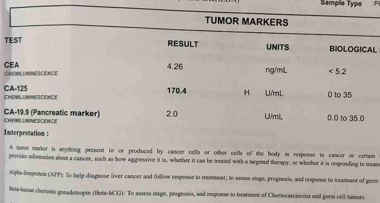
Therefore, CA-125 should always be interpreted together with clinical examination, ultrasound, CT/MRI findings and histopathology rather than in isolation.
Can a 19 cm Ovarian Tumour Be Removed Safely When It Is Touching the Bladder, Bowel and Ureter?
Large ovarian tumours often appear intimidating on CT scans because they may compress several important organs. However, compression does not always mean invasion.
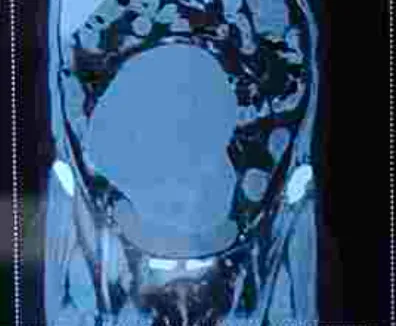
In one patient treated at the Indian Ovarian Cancer Institute (IOCI), Ahmedabad, CT scan demonstrated a 12 × 15.7 × 19 cm complex ovarian mass abutting the urinary bladder, adjacent bowel loops, sigmoid colon and anterior abdominal wall. The tumour also caused mild compression of the right ureter with resulting hydroureteronephrosis. Importantly, there was no evidence of invasion into major blood vessels. During surgery, meticulous retroperitoneal dissection and adhesiolysis allowed careful separation of the tumour from surrounding structures before comprehensive staging surgery was completed.
- Surgical Perspective
Unlike many gastrointestinal cancers, ovarian tumours often displace surrounding organs rather than infiltrate them directly. Large tumour size and expected adhesions usually make open surgery the preferred approach, allowing safe identification of the ureter, bowel and major blood vessels while maximizing the chance of complete tumour removal according to oncologic principles.
Why Were 35 Lymph Nodes Removed Even Though the CT Scan Did Not Show Enlarged Lymph Nodes?
Modern CT scans are excellent for evaluating ovarian cancer, but they cannot detect every microscopic tumour deposit. This is why comprehensive surgical staging remains an important part of ovarian cancer treatment.
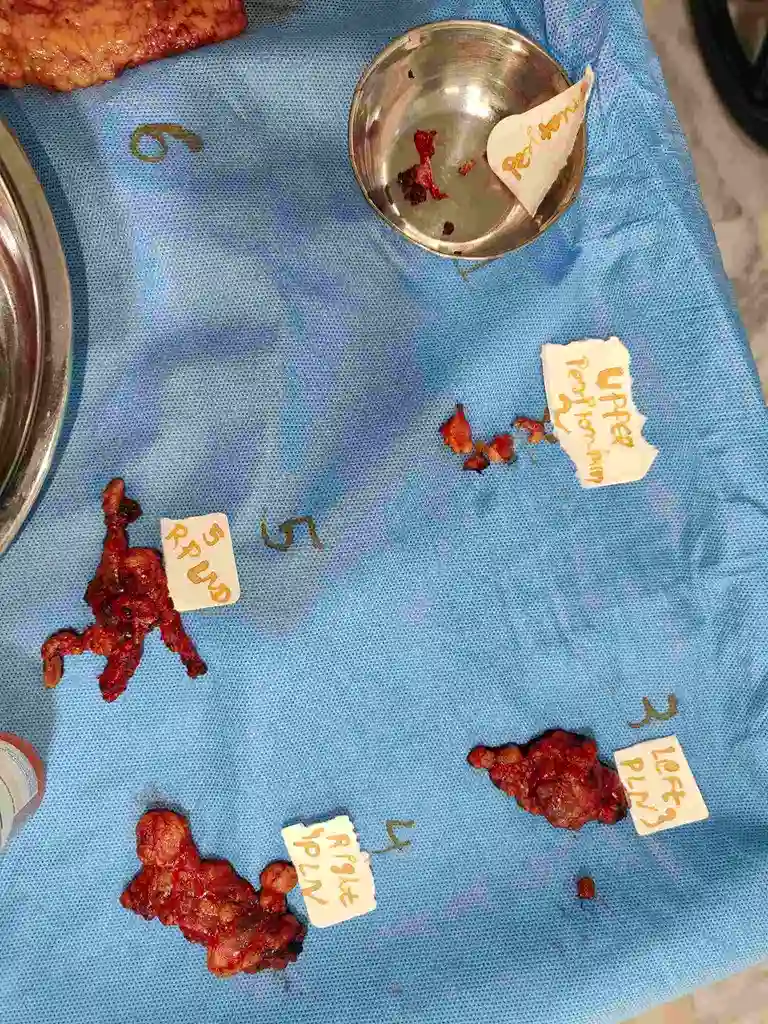
In this case managed at the Indian Ovarian Cancer Institute (IOCI), Ahmedabad, preoperative CT showed no significant lymphadenopathy, no liver metastasis and no radiological evidence of peritoneal carcinomatosis. However, after frozen section confirmed ovarian malignancy, comprehensive staging surgery included bilateral pelvic and retroperitoneal lymph node dissection. Final histopathology revealed one microscopic metastatic lymph node among 35 nodes examined, changing the final diagnosis to FIGO Stage IIIA1(i) despite reassuring imaging findings.
- Clinical Insight
CT and MRI identify enlarged lymph nodes but may miss microscopic metastases. Surgical staging provides the most accurate assessment of disease spread, helps determine the correct FIGO stage and guides recommendations for postoperative chemotherapy and long-term follow-up.
Fallopian Tube Cancer and Ovarian Cancer Are Treated in a Similar Way
A recently managed case at the Indian Ovarian Cancer Institute (IOCI), Ahmedabad, highlights an important fact—every cancer that appears to be an ovarian cancer on imaging does not necessarily arise from the ovary.
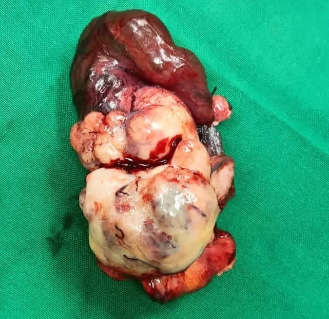
⦿ MRI and CT showed a malignant left adnexal mass closely related to the ovary with associated hydrosalpinx, suggesting an ovarian malignancy. During surgery, the tumour was removed intact and comprehensive staging surgery was performed. Final histopathology showed that the primary tumour originated from the left fallopian tube (High-Grade Serous Carcinoma), while both ovaries were free of tumour.
⦿ Today, it is well recognized that many cancers traditionally labelled as “ovarian cancer,” especially high-grade serous carcinoma, actually begin in the fallopian tube.
⦿ Fortunately, the treatment principles for epithelial ovarian cancer and fallopian tube cancer are essentially the same. Complete surgical staging—including assessment of pelvic and retroperitoneal lymph nodes, omentum and peritoneum—is important to accurately determine disease extent and guide further treatment.
Accurate pathology after surgery is therefore essential, as it establishes the true site of origin while ensuring appropriate management.
CT Scan Suggested Omental Disease—Final Histopathology Told a Different Story
A recently managed ovarian/fallopian tube cancer case at the Indian Ovarian Cancer Institute (IOCI), Ahmedabad, demonstrates why CT scan findings must always be interpreted together with surgical findings and histopathology.
The preoperative CT scan showed a solid-cystic pelvic tumour with surrounding peritoneal thickening and omental fat stranding, findings that raised suspicion of omental involvement by ovarian cancer.
During surgery, the omentum and suspicious peritoneal areas were carefully evaluated and removed as part of complete staging surgery.
- However, final histopathology showed:
⦿ Omentum – Negative for tumour
⦿ Pelvic peritoneal biopsy – Negative
⦿ Upper peritoneal biopsy – Negative
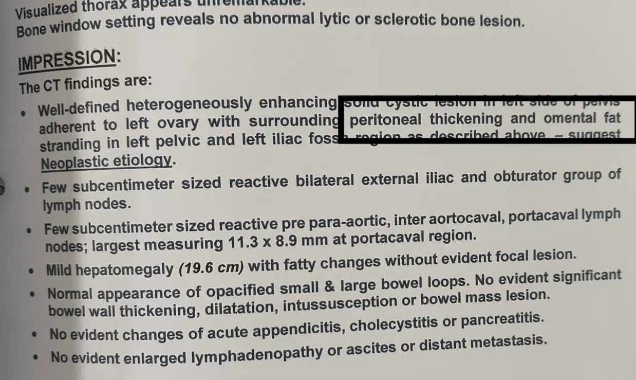
This illustrates an important concept in ovarian cancer care. CT scan is excellent for identifying areas that require careful surgical evaluation, but imaging alone cannot always distinguish metastatic disease from inflammatory or reactive changes.
The final stage of ovarian or fallopian tube cancer should always be based on surgical findings and histopathological confirmation rather than imaging alone.
Sentinel Lymph Node in Ovarian & Fallopian Tube Cancer – Is It Ready for Routine Practice?
Sentinel lymph node biopsy has become standard in endometrial cancer, but its role in ovarian and fallopian tube cancers is still evolving.
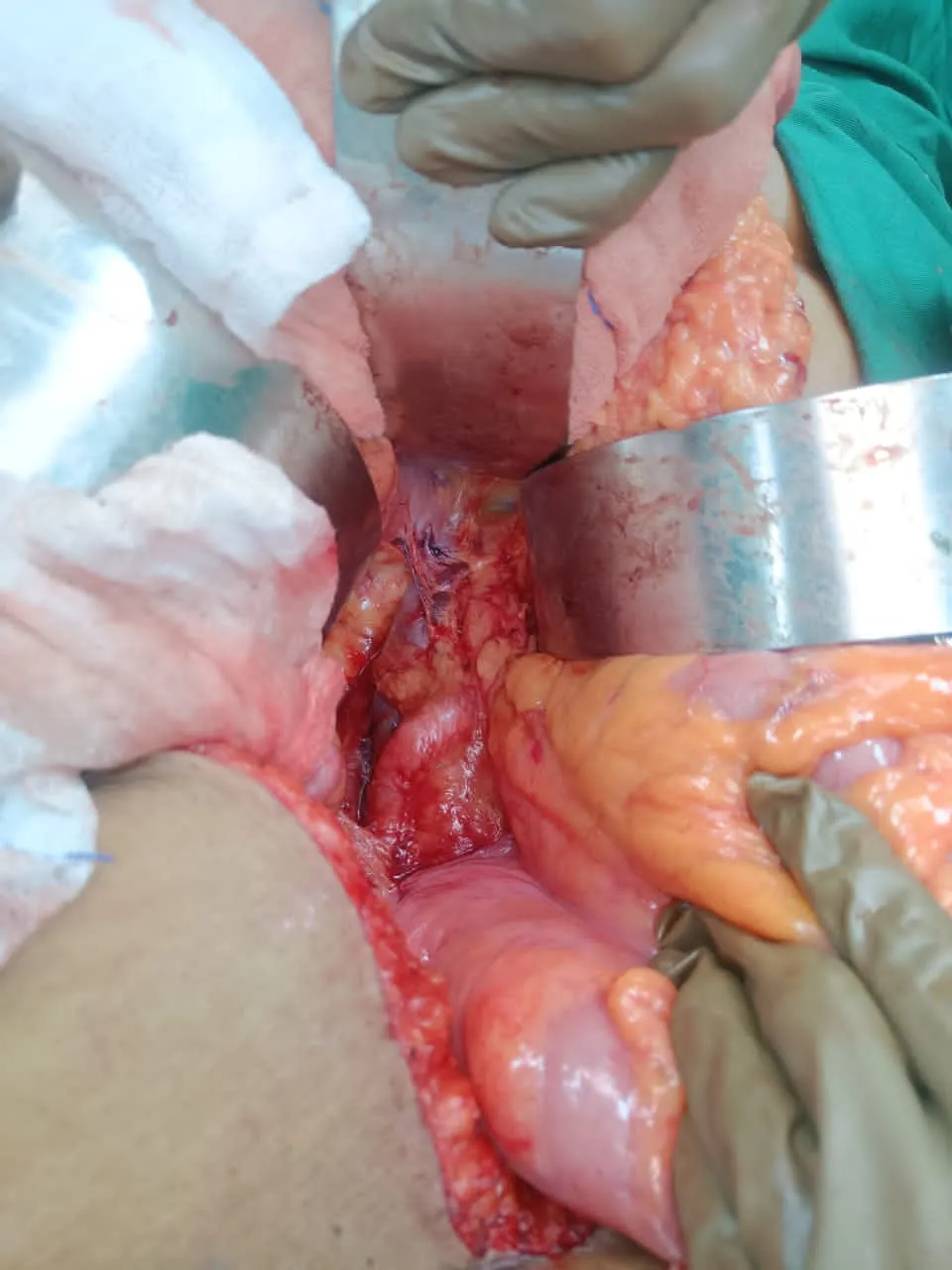
- In this case, intraoperative sentinel retroperitoneal lymph nodes were identified along with complete pelvic and retroperitoneal lymph node dissection . Final pathology showed:
⦿ Sentinel nodes – Negative
⦿ Pelvic nodes – Negative
⦿ Retroperitoneal nodes – Negative
A total of 43 lymph nodes were examined, and all were free of metastasis.
Because sentinel node mapping is still under evaluation in ovarian/fallopian tube cancer, complete staging lymphadenectomy remains important in appropriately selected patients.
Future research may reduce the need for extensive nodal dissection in some women, but until then, accurate staging remains essential for treatment planning.
Recovery After Open Ovarian Cancer Surgery Can Begin Earlier Than Many Patients Expect
Many patients worry that recovery after open ovarian cancer surgery will require prolonged bed rest and several weeks before they can move comfortably.
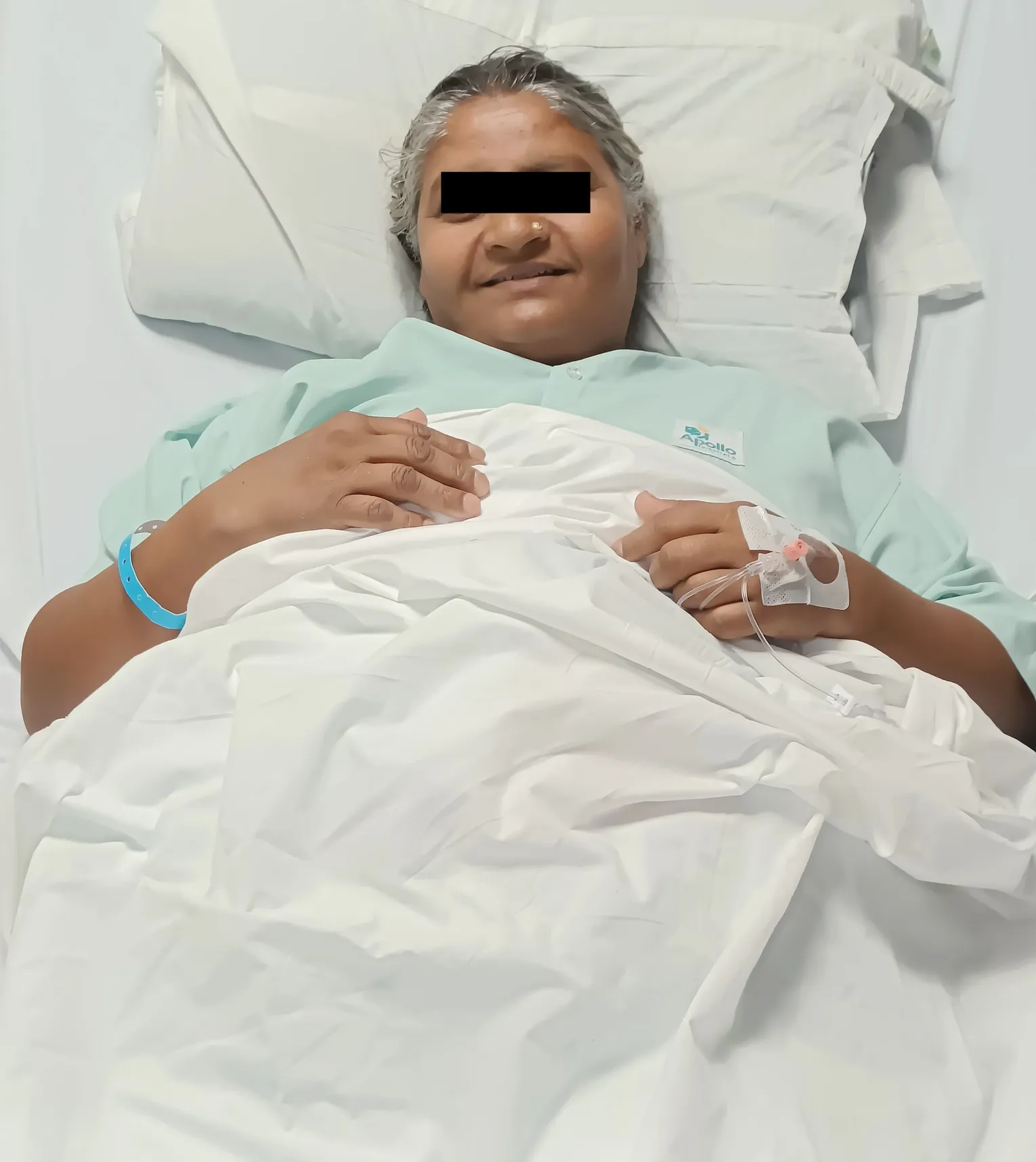
⦿ A recently managed case at the Indian Ovarian Cancer Institute (IOCI), Ahmedabad, demonstrates how modern perioperative care helps patients recover earlier even after extensive abdominal surgery.
⦿ The patient underwent open surgery that included tumour removal, hysterectomy, bilateral salpingo-oophorectomy, pelvic and retroperitoneal lymph node dissection, omentectomy and peritoneal biopsies.
⦿ With structured postoperative care, good pain control, early nutrition, physiotherapy, breathing exercises and early mobilisation (ERAS – Enhanced Recovery After Surgery principles), the postoperative course remained smooth. The patient became ambulatory early, remained clinically stable and was discharged within five days.
⦿ For many women undergoing ovarian cancer surgery, one of the most common questions is, “When will I be able to walk, eat normally and return to daily activities?” In appropriately selected patients, early walking with adequate medical support not only improves confidence but also reduces complications such as blood clots, chest infections and delayed bowel recovery.
⦿ Recovery after ovarian cancer surgery is a step-by-step process, and modern ERAS protocols are designed to help patients regain independence as safely and as early as possible.
Get health updates from Dr Swati Shah on WhatsApp
Swasth Parivar: twice a month, practical advice on recovery, diet and warning signs. No advertising. Stop any time.
Your number is used for these updates and for the clinic to reach you. It is never sold or passed on. To stop, reply STOP on WhatsApp.