


Case Library - Ovary Cyst/Tumour surgery
Can Fertility Be Preserved in Young Girls with Ovarian Cancer? How Frozen Section Helps Surgeons Decide
Preserving future fertility is an important consideration when treating ovarian tumours in children, adolescents and young women. However, fertility-preserving surgery (FSS) is appropriate only in carefully selected patients. The decision depends on the type of ovarian tumour, the extent of disease and accurate surgical staging.
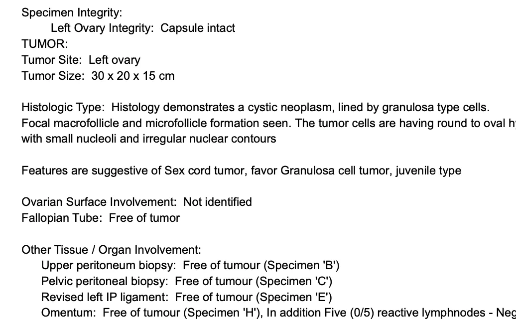
⦿ In this 12-year-old girl treated at the Indian Ovarian Cancer Institute (IOCI), Ahmedabad, the ovarian tumour was removed intact and immediately examined by frozen section, a rapid pathological assessment performed while the patient remained under anaesthesia. Frozen section suggested a malignant sex cord stromal tumour, allowing the surgical team to confidently proceed with fertility-preserving surgery by preserving the uterus and the opposite ovary while completing comprehensive surgical staging, including peritoneal biopsies, omentectomy, pelvic and retroperitoneal lymph node dissection. Final histopathology confirmed a Juvenile Granulosa Cell Tumour confined to the left ovary (FIGO Stage IA) with negative peritoneal biopsies, omentum and 41 negative lymph nodes.
- Expert Insight
Fertility-preserving surgery is not suitable for every ovarian cancer. It is considered only when the tumour type and stage indicate that preserving the uterus and the opposite ovary is oncologically safe. Frozen section provides the intraoperative diagnosis that helps surgeons make this decision immediately, allowing both appropriate cancer surgery and preservation of future fertility whenever medically feasible.
Can Fertility Be Preserved in Young Girls with Ovarian Cancer? The Answer Depends on the Tumour Type, Stage and Safe Removal of the Tumour
⦿ For children, adolescents and young women diagnosed with an ovarian tumour, one of the biggest concerns is whether future fertility can be preserved. The answer depends on three important factors: the type of tumour, the stage of disease, and whether the tumour can be removed intact without rupture.
⦿ A 12-year-old girl recently treated at the Indian Ovarian Cancer Institute (IOCI), Ahmedabad had a 30 × 20 × 15 cm left ovarian tumour. The mass was carefully removed intact, followed by frozen section (“live biopsy”), which suggested a malignant sex cord stromal tumour. This intraoperative diagnosis allowed the surgical team to perform fertility-preserving surgery, preserving the uterus and the opposite ovary while completing comprehensive surgical staging with peritoneal biopsies, omentectomy and pelvic plus retroperitoneal lymph node dissection. Final histopathology confirmed a Juvenile Granulosa Cell Tumour, FIGO Stage IA, with intact capsule, negative washings, negative peritoneal biopsies and 41 negative lymph nodes.
- Guideline Insight
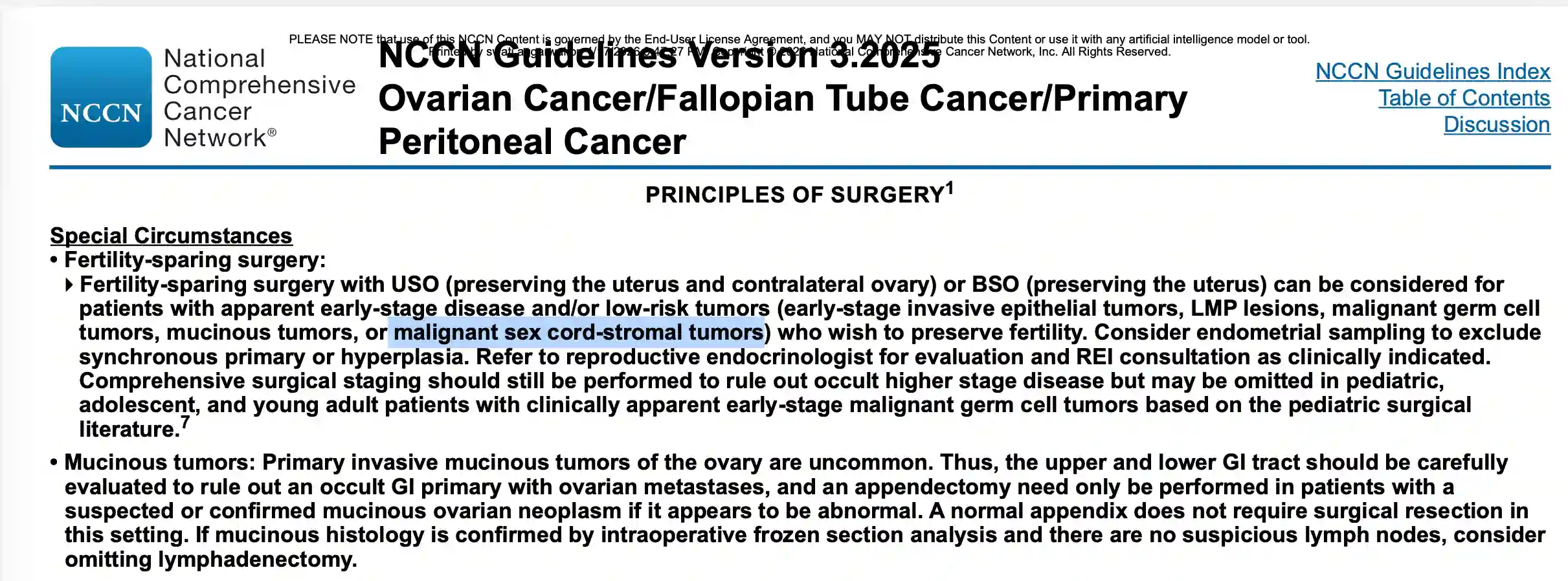
Current NCCN Guidelines state that fertility-sparing surgery can be considered for selected patients with apparent early-stage, low-risk ovarian tumours, including malignant sex cord-stromal tumours, while still performing appropriate surgical staging. Preserving fertility is therefore not simply a matter of age—it is possible only when the tumour biology, stage and oncologic principles make it safe. Removing the tumour without rupture is especially important because intraoperative spill may upstage an otherwise Stage IA tumour to Stage IC in appropriate circumstances, which can influence recommendations regarding adjuvant chemotherapy. Careful preoperative planning and meticulous surgical technique are therefore essential to achieve both cancer control and fertility preservation whenever medically appropriate.
Why Are Retroperitoneal Lymph Nodes So Important in Ovarian Cancer Surgery?
When ovarian cancer spreads to lymph nodes, many people assume it first involves the pelvic lymph nodes. However, the ovaries primarily drain through the ovarian blood vessels to the para-aortic (retroperitoneal) lymph nodes, making these nodes an important part of surgical staging.
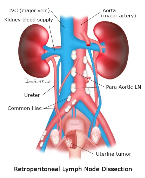
⦿ In one patient recently treated at the Indian Ovarian Cancer Institute (IOCI), Ahmedabad, imaging did not show enlarged lymph nodes. During surgery, comprehensive staging included both bilateral pelvic lymph node dissection and retroperitoneal (para-aortic) lymph node dissection after frozen section confirmed high-grade ovarian cancer. Final histopathology demonstrated a microscopic metastasis in one retroperitoneal lymph node, while all 22 pelvic lymph nodes were negative. This changed the final stage to FIGO Stage IIIA1(i) and influenced further treatment planning.
- Clinical Insight
Retroperitoneal lymph node dissection is one of the most technically demanding steps in ovarian cancer surgery because these lymph nodes lie close to the aorta, inferior vena cava, ureters and major abdominal blood vessels. This case illustrates why ovarian cancer surgery requires an understanding of the natural lymphatic drainage of the ovary, not just removal of the pelvic tumour. Careful staging can identify microscopic disease that may not be visible on CT or MRI and ensures patients receive the most appropriate postoperative treatment.
Weekly Chemotherapy for Elderly or Frail Patients with Advanced Ovarian Cancer: An Effective Neoadjuvant Approach
A recently managed case at the Indian Ovarian Cancer Institute (IOCI), Ahmedabad involved a 73-year-old woman with Stage IIIC/advanced high-grade serous ovarian cancer, poor performance status, multiple co-morbidities and extensive peritoneal disease. After evaluation by a Gynecologic Oncologist, Neoadjuvant Chemotherapy with 9 weekly cycles of Paclitaxel + Carboplatin was chosen before Cytoreductive Surgery. The patient subsequently underwent successful interval Cytoreductive Surgery after good clinical and radiological response.
- Clinical Insight
Many patients ask:
⦿ Is chemotherapy safe after 70 years of age?
⦿ Can weak patients tolerate ovarian cancer chemotherapy?
⦿ Is weekly chemotherapy better than 3-weekly chemotherapy?
⦿ Can elderly patients still undergo ovarian cancer surgery later?
For carefully selected patients, weekly Paclitaxel + Carboplatin may be considered as Neoadjuvant Chemotherapy (NACT), especially when age, nutritional status, co-morbidities or performance status make immediate major surgery unsafe.
- The goals are to:
⦿ Improve performance status
⦿ Reduce tumour burden
⦿ Improve nutritional status
⦿ Optimise medical fitness
⦿ Increase the chances of successful Cytoreductive Surgery
Treatment planning should always be individualised by a Gynecologic Oncologist based on disease extent, fitness and response to therapy.
Take-home message:
Advanced age alone is not a contraindication for ovarian cancer treatment. With appropriate patient selection, weekly Neoadjuvant Chemotherapy, multidisciplinary optimisation and treatment at an experienced ovarian cancer centre, even elderly patients can safely proceed to successful Cytoreductive Surgery.
Which Ultrasound Findings in an Ovarian Cyst Require Further Evaluation?
Many ovarian cysts are harmless and disappear on their own, but some ultrasound findings require further assessment by a gynecologic oncologist.
⦿ A recently managed patient at the Indian Ovarian Cancer Institute (IOCI), Ahmedabad had ultrasound showing a large 21 cm multiloculated abdominopelvic cystic mass with numerous thin septations and low-level internal echoes. Although there was no ascites or enlarged lymph nodes, the lesion was large and had complex features, prompting CT evaluation and surgery. Final histopathology confirmed a Benign Mucinous Cystadenoma with an intact capsule.
- Clinical Insight
An ovarian cyst should undergo further evaluation when ultrasound shows one or more of the following:
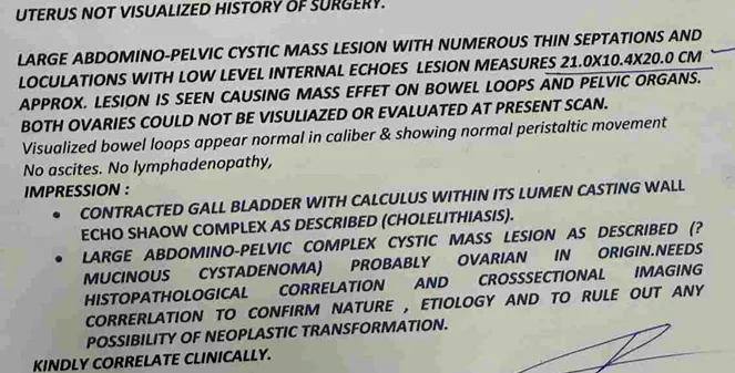
⦿ Large size (especially >10 cm)
⦿ Multiple septations
⦿ Solid components or papillary projections
⦿ Internal vascularity on Doppler
⦿ Ascites
⦿ Bilateral ovarian masses
⦿ Enlarged lymph nodes
⦿ These findings do not always mean cancer, but they indicate that additional imaging, tumour markers and specialist evaluation may be needed.
Can CT or Ultrasound Tell Whether an Ovarian Tumour Is Benign or Malignant?
Imaging plays a vital role in evaluating ovarian masses, but it cannot always provide a definite diagnosis. In this patient treated at the Indian Ovarian Cancer Institute (IOCI), Ahmedabad, CT showed a 24 cm multiseptated pelvic mass with enhancing septations that displaced the bowel loops, bladder, ureters and major blood vessels. Fortunately, there was no ascites, liver metastasis, bulky lymph nodes or obvious invasion of surrounding structures. Despite these reassuring features, imaging could not confidently exclude a borderline or malignant mucinous tumour. Final histopathology demonstrated a Benign Mucinous Cystadenoma.
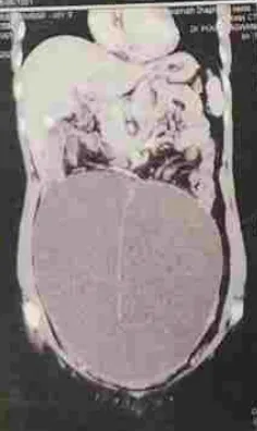
Clinical Insight
- Imaging features that favour a benign ovarian cyst include:
⦿ Thin, smooth wall
⦿ Simple fluid content
⦿ No solid nodules
⦿ Minimal or no internal blood flow
⦿ No ascites or enlarged lymph nodes
- Features that raise suspicion for malignancy include:
⦿ Thick or irregular septations
⦿ Solid or papillary components
⦿ Internal vascularity
⦿ Omental deposits or enlarged lymph nodes
⦿ Spread beyond the ovary
Imaging estimates the risk of malignancy—it does not replace histopathology.
Which Tumour Markers Should Be Tested for an Ovarian Mass? The Answer Depends on the Patient's Age
Many patients believe CA-125 is the only blood test needed for an ovarian tumour. In reality, the choice of tumour markers depends on the patient’s age and the suspected tumour type.
⦿ A recently managed patient at the Indian Ovarian Cancer Institute (IOCI), Ahmedabad had a 24 cm ovarian mass, while CA-125 (19.78 U/mL) and CA19-9 (<2 U/mL) were not significantly elevated. Surgery was still recommended because imaging suggested a large complex ovarian neoplasm. Final histopathology confirmed a Benign Mucinous Cystadenoma.
- Clinical Insight
Different ovarian tumours produce different tumour markers. Depending on the clinical scenario, doctors may request:
⦿ CA-125 – Mainly epithelial ovarian tumours
⦿ HE4 – May improve risk assessment in selected epithelial tumours
⦿ CEA & CA19-9 – Helpful when mucinous ovarian tumours are suspected
⦿ AFP, β-hCG & LDH – Important in children and younger women when germ cell tumours are possible
⦿ Inhibin A/B – Suggested when a sex cord-stromal tumour (such as granulosa cell tumour) is suspected
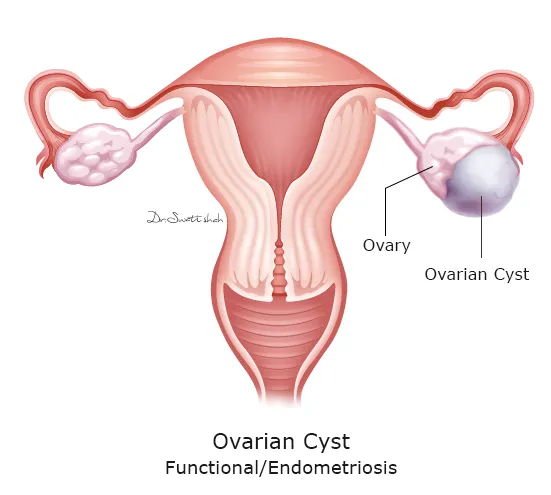
Tumour markers support diagnosis but cannot confirm or exclude ovarian cancer on their own. They must always be interpreted together with imaging and clinical findings.
When Does an Ovarian Cyst Need Surgery? Why Is a Biopsy Usually Avoided?
⦿ In this patient managed at the Indian Ovarian Cancer Institute (IOCI), Ahmedabad, imaging showed a 24 cm complex ovarian tumour. Because of its size and suspicious imaging features, surgery was indicated. The tumour was removed intact without rupture, and frozen section performed during surgery confirmed a mucinous cystadenoma, allowing the surgical team to make informed intraoperative decisions. Final histopathology confirmed a Benign Mucinous Cystadenoma with no borderline tumour or malignancy.
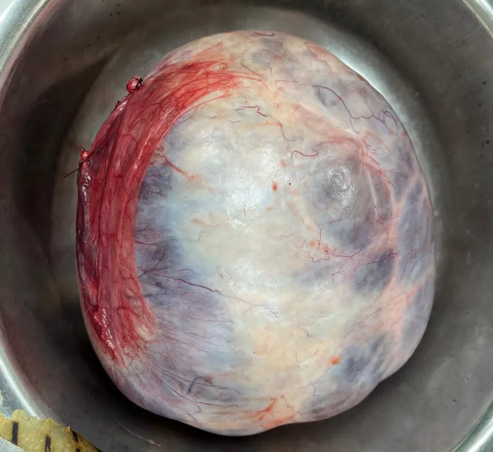
Clinical Insight
- Surgery is generally recommended for ovarian cysts that are:
⦿ Large (commonly >5–10 cm, depending on age and imaging findings)
⦿ Complex or suspicious on imaging
⦿ Symptomatic
⦿ Increasing in size
⦿ Associated with concerning tumour markers or an uncertain diagnosis
- When surgery is needed, removing the tumour intact and using frozen section often provides the safest and most accurate approach for diagnosis and treatment planning.
Can MRI Reliably Differentiate a Borderline Ovarian Tumour from a Benign Ovarian Cyst or Ovarian Cancer?
Recently managed at the Indian Ovarian Cancer Institute (IOCI), Ahmedabad, this patient had MRI showing a large multiloculated right ovarian cyst with internal septal thickening. The radiological differential diagnosis included mucinous cystadenoma, borderline ovarian tumour and tubo-ovarian pathology, highlighting the difficulty in establishing a definitive diagnosis before surgery.
- Clinical Insight
Borderline ovarian tumours often have imaging features that overlap with both benign ovarian cysts and invasive ovarian cancer.
MRI and CT may raise suspicion when they show:
⦿ Multiloculated cysts
⦿ Thick septations
⦿ Papillary projections
⦿ Mural nodules
⦿ Proteinaceous or mucinous contents
⦿ Solid enhancing areas
However, no imaging modality can reliably distinguish a benign ovarian tumour, borderline ovarian tumour and ovarian cancer in every patient.
Definitive diagnosis requires complete surgical removal of the tumour followed by detailed histopathological examination.
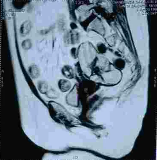
Take-home message:
Imaging helps estimate the likelihood of malignancy, but the final diagnosis depends on pathology after surgery.
Why Is It So Important Not to Rupture a Borderline Ovarian Tumour During Surgery?
⦿ A recently operated case at the Indian Ovarian Cancer Institute (IOCI), Ahmedabad highlights one of the most important surgical principles in managing borderline ovarian tumours.
⦿ Unlike a simple ovarian cyst, a borderline ovarian tumour should ideally be removed intact without rupture.
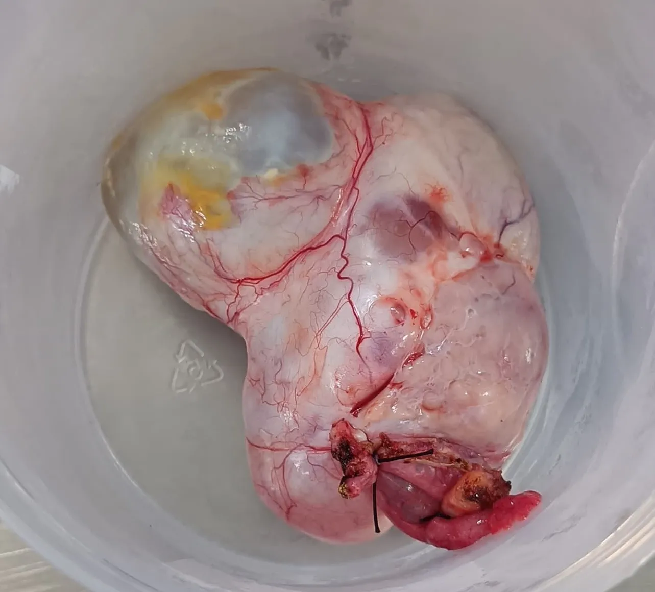
Clinical Insight
- Although borderline ovarian tumours are not invasive ovarian cancers, they are also not completely benign. For this reason:
⦿ Preoperative needle biopsy or cyst aspiration is generally avoided.
⦿ Tumour rupture during surgery should be prevented whenever possible.
⦿ Spillage of tumour cells into the abdominal cavity may increase the risk of peritoneal implants and local recurrence.
⦿ Chemotherapy has a limited role in treating borderline ovarian tumours, making optimal surgery the cornerstone of treatment.
⦿ Removing the tumour intact allows accurate histopathological evaluation while reducing the chances of disease recurrence related to tumour spillage.
Take-home message:
For borderline ovarian tumours, meticulous surgery with intact tumour removal is one of the most important factors influencing long-term outcomes.
Absent Kidney Detected During Ovarian Tumour Evaluation—Why Does It Matter Before Surgery?
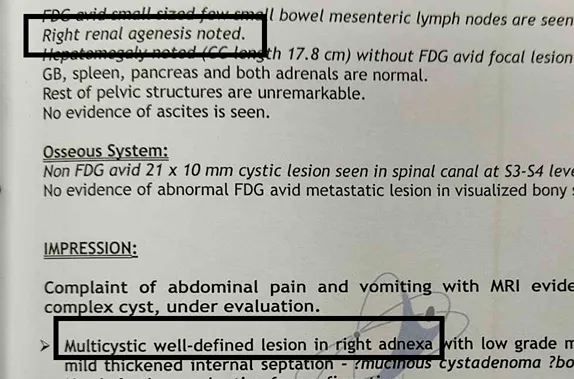
During the evaluation of a recent ovarian tumour case at the Indian Ovarian Cancer Institute (IOCI), Ahmedabad, MRI revealed an important incidental finding—the patient had congenital absence of the right kidney (renal agenesis) with compensatory enlargement of the left kidney.
Although unrelated to the ovarian tumour itself, this information was highly relevant for surgical planning.
- Clinical Insight
Before pelvic surgery, surgeons carefully review imaging not only to assess the tumour but also to identify anatomical variations that may influence the operation.
Examples include:
⦿ Congenital absent kidney
⦿ Ectopic pelvic kidney
⦿ Duplicated ureter
⦿ Aberrant blood vessels
Such variations can alter the normal course of the ureter and surrounding vascular anatomy. Recognizing these findings before surgery helps prevent inadvertent injury and allows safer pelvic dissection.
Take-home message:
Preoperative imaging provides much more than tumour diagnosis—it also helps surgeons understand individual anatomy and perform safer ovarian tumour surgery.
Why Is CA 19-9 Tested Along with CA-125 in Some Ovarian Cysts and Ovarian Tumours?
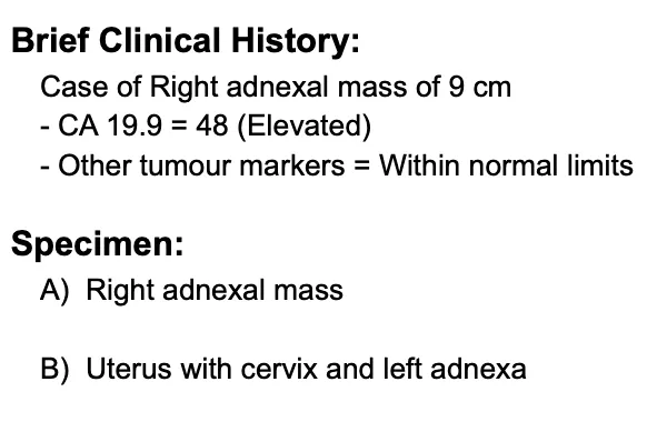
A recently managed ovarian tumour at the Indian Ovarian Cancer Institute (IOCI), Ahmedabad highlights why evaluation of a complex ovarian mass often requires more than a single tumour marker.
While CA-125 is the most widely recognized marker for ovarian cancer, CA 19-9 can provide valuable additional information in selected patients.
Clinical Insight
- CA 19-9 may be elevated in:
⦿ Mucinous ovarian tumours
⦿ Borderline mucinous ovarian tumours
⦿ Some epithelial ovarian cancers
⦿ Endometriosis
⦿ Mature cystic teratoma
- It may also be raised in conditions outside the ovary, including:
⦿ Pancreatic cancer
⦿ Benign hepatobiliary diseases
⦿ Pancreatitis
For this reason, doctors often interpret CA-125, CA 19-9, CEA and other age-appropriate tumour markers together with imaging findings. If CA 19-9 is elevated before surgery, it can also serve as a useful baseline marker during postoperative follow-up.
Take-home message:
No single tumour marker can diagnose an ovarian tumour. Combining tumour markers with imaging and histopathology provides the most accurate assessment and helps guide long-term follow-up.
CT Suggested a Dermoid Cyst. Frozen Section Changed the Entire Surgery.
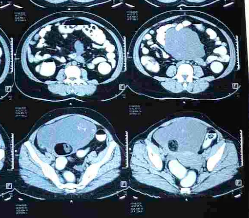
A 44-year-old woman presented with only 3–4 days of abdominal heaviness and increased urinary frequency. CT abdomen demonstrated a large 16.3 × 11.8 × 18 cm well-defined right adnexal solid-cystic mass with internal fat density, tiny calcific foci and enhancing solid components, making it highly suggestive of a mature cystic teratoma (dermoid cyst). There was no significant abdominopelvic lymphadenopathy, no bowel or peritoneal abnormality and only minimal reactive free fluid. Tumour markers were also not typical of epithelial ovarian cancer (CA-125 37.4 U/ml).
⦿ Because the diagnosis became available during the same operation, the procedure was immediately extended to include total hysterectomy, contralateral salpingo-oophorectomy, bilateral pelvic lymph node dissection, retroperitoneal lymphadenectomy and supracolic omentectomy, avoiding a second major surgery. Final histopathology confirmed a mucinous borderline tumour (intestinal type) associated with a central mature cystic teratoma (dermoid cyst).
- Clinical Insight
When managing any large ovarian cyst, tumour or complex adnexal mass, surgery should be planned with the possibility of unexpected pathology in mind. Frozen section can immediately alter surgical management and allow complete oncologic treatment during the same anaesthesia.
Indian Ovarian Cancer Institute (IOCI), Ahmedabad
Every patient is unique. Treatment recommendations are individualized based on clinical evaluation, imaging, pathology and multidisciplinary discussion.
Are All Dermoid (Teratoma) Cysts Benign? This Case Demonstrates Why Histopathology Matters.
CT imaging demonstrated features classically associated with a mature cystic teratoma—a well-defined solid-cystic ovarian lesion containing macroscopic fat, calcification and heterogeneous enhancing solid components, inseparable from the right ovary. These imaging characteristics are considered highly characteristic of a dermoid cyst.
However, imaging predicts the most likely diagnosis, not the final diagnosis.
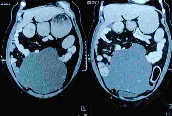
Microscopic examination of the resected specimen demonstrated a 20.5 × 16 × 8 cm mucinous borderline tumour (intestinal type) with a central mature cystic teratoma (dermoid cyst). The cut surface showed multiloculated cysts filled with mucinous material, while a smaller cystic area contained cheesy material with hair follicles, explaining why the radiological appearance closely resembled a dermoid.
This case illustrates that ovarian tumours may contain more than one histological component. The presence of dermoid elements on imaging does not completely exclude coexisting borderline or neoplastic pathology.
- Clinical Insight
Radiology guides surgical planning, but histopathology remains the definitive diagnosis. Large or complex ovarian masses should therefore be removed using oncologic principles, even when imaging appears reassuring.
Indian Ovarian Cancer Institute (IOCI), Ahmedabad
Every patient is unique. Treatment recommendations are individualized based on clinical evaluation, imaging, pathology and multidisciplinary discussion.
Why Frozen Section Allowed Complete Staging of This Borderline Ovarian Tumour in One Operation
During surgery, the right ovarian mass measured approximately 18 × 16 cm and was associated with mucinous free fluid in the pelvis. The upper abdomen, appendix and colon appeared grossly normal, with no evidence of disease elsewhere. Instead of completing only ovarian cyst surgery, the specimen was immediately examined using frozen section biopsy.
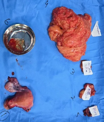
⦿ Frozen section identified a mucinous borderline ovarian tumour, allowing the surgical plan to change instantly. During the same operation, comprehensive surgical staging was completed, including total hysterectomy, bilateral salpingectomy, left oophorectomy, bilateral pelvic lymph node dissection, retroperitoneal lymphadenectomy and supracolic omentectomy.
⦿ Final pathology demonstrated no ovarian surface involvement, no fallopian tube involvement, no implants, negative omentum, and all 32 lymph nodes free of tumour (0/32), resulting in a pathological stage of pT1c2N0.
- Clinical Insight
Borderline ovarian tumours are biologically different from invasive ovarian cancer, but accurate surgical staging remains essential. Frozen section helps avoid under-treatment and often prevents the need for a second staging operation after the final pathology report becomes available.
Indian Ovarian Cancer Institute (IOCI), Ahmedabad
Every patient is unique. Treatment recommendations are individualized based on clinical evaluation, imaging, pathology and multidisciplinary discussion.
A Nearly 18 cm Ovarian Tumour Caused Only Mild Symptoms—Even Though the Patient Was a Doctor
One of the most striking aspects of this case was not the pathology—it was the presentation.
Despite being a medical professional, the patient experienced only 3–4 days of abdominal heaviness and increased urinary frequency before evaluation. Imaging then revealed a mass measuring 16.3 × 11.8 × 18 cm (approximately 1792 cm³) occupying the right adnexa.
The CT scan demonstrated a large solid-cystic lesion containing fat density, calcification and enhancing components, while the remainder of the abdomen showed no bowel wall thickening, no peritoneal thickening, no significant lymphadenopathy and only minimal reactive free fluid.

This reflects a common characteristic of ovarian tumours. Because the ovaries are located within the abdominal cavity, masses can enlarge considerably before producing obvious symptoms. Many women present only when the tumour causes pressure on surrounding organs, abdominal fullness or urinary symptoms.
- Clinical Insight
The severity of symptoms does not always reflect the size or biological behaviour of an ovarian tumour. Persistent abdominal enlargement, pelvic heaviness or a newly detected ovarian mass should be evaluated carefully, even when discomfort is minimal.
Indian Ovarian Cancer Institute (IOCI), Ahmedabad
Every patient is unique. Treatment recommendations are individualized based on clinical evaluation, imaging, pathology and multidisciplinary discussion.
Pleural Effusion in an Ovarian Tumour Is Not Always Stage IV Cancer
One important lesson in gynecologic oncology is that pleural effusion (fluid around the lungs) does not automatically mean advanced metastatic ovarian cancer.
⦿ In this case, imaging showed a large left ovarian tumour, mild ascites, and bilateral pleural effusion, which could easily have suggested advanced disease. Instead of assuming metastasis, the pleural fluid was examined through cytology, and no malignant cells were identified. This changed the entire treatment pathway.
⦿ The patient underwent fertility-preserving surgery with intact removal of the ovarian tumour, along with comprehensive surgical staging including peritoneal biopsies, omentectomy, lymph node assessment, and fluid cytology. Final histopathology confirmed a dysgerminoma confined to the left ovary (pT1N0), with no tumour in the peritoneum, omentum, ascitic fluid, or regional lymph nodes.
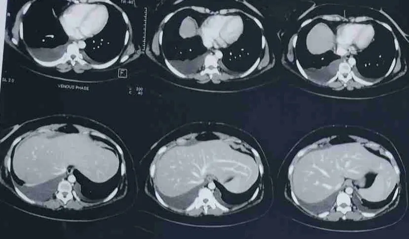
This meant the disease was Stage I, not Stage IV, and the patient could avoid unnecessary chemotherapy while preserving future fertility.
- Clinical Insight
In ovarian tumours, pleural effusion may be reactionary or inflammatory rather than metastatic. Cytological confirmation is essential before assigning advanced-stage disease and planning treatment.
Indian Ovarian Cancer Institute (IOCI), Ahmedabad
Every patient is unique. Treatment recommendations are individualized based on clinical evaluation, imaging, pathology and multidisciplinary discussion.
Why Fertility-Preserving Surgery Was Possible in bilateral Ovarian Tumour
A common concern for young women with an ovarian tumour is whether treatment will affect future fertility.
In this case, a large left ovarian germ cell tumour was identified, while the opposite ovary contained a small cystic lesion. Rather than performing surgery that removed both ovaries, a careful fertility-preserving approach was chosen.
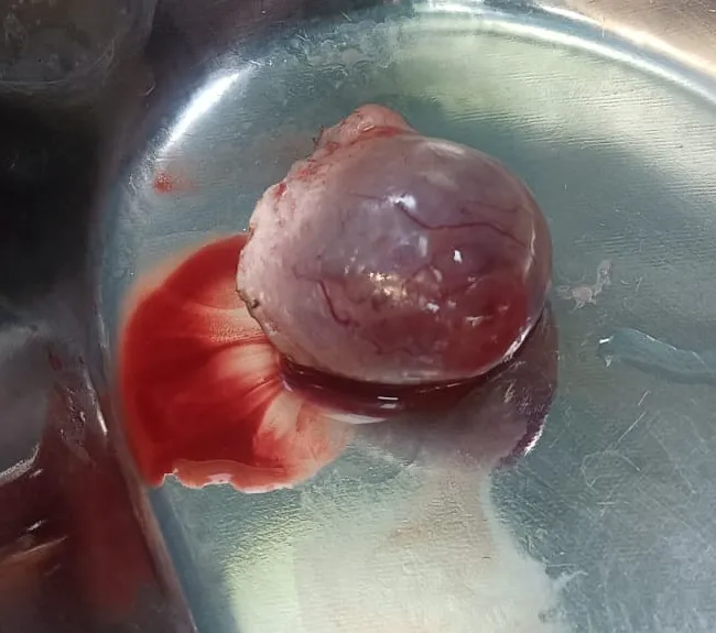
The left ovarian tumour was removed intact with left salpingo-oophorectomy, while the right ovarian cyst was excised and the right ovary was preserved. Intraoperative frozen section and final histopathology confirmed that the left-sided tumour was dysgerminoma, whereas the right ovarian lesion was a benign haemorrhagic luteal/follicular cyst. Comprehensive surgical staging showed no spread to the peritoneum, omentum, ascitic fluid, or lymph nodes, supporting Stage I disease.
Because the tumour was confined to one ovary and complete staging was performed, fertility preservation was oncologically appropriate.
- Clinical Insight
Fertility-preserving surgery can be considered in selected young women with ovarian germ cell tumours or certain ovarian cysts/tumours, particularly when disease appears limited to one ovary and proper surgical staging is performed.
Indian Ovarian Cancer Institute (IOCI), Ahmedabad
Every patient is unique. Treatment recommendations are individualized based on clinical evaluation, imaging, pathology and multidisciplinary discussion.
How Comprehensive Surgical Staging Can Prevent Unnecessary Chemotherapy
In ovarian tumours, surgery is often performed not only to remove the tumour but also to accurately determine the stage of disease.
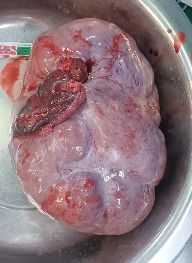
⦿ In this case, the patient underwent exploratory laparotomy with intact removal of the left ovarian tumour, right ovarian cyst excision, peritoneal biopsies, supracolic omentectomy, bilateral pelvic lymph node dissection, retroperitoneal lymph node dissection, and fluid cytology.
⦿ Although preoperative imaging suggested ascites, pleural effusion, and possible peritoneal involvement, the staging procedure demonstrated a very different picture. Peritoneal biopsies were tumour-free, the omentum was free of tumour, ascitic/fluid cytology showed no malignant cells, and all 37 regional lymph nodes were negative for metastasis.
⦿ This comprehensive assessment established Stage I dysgerminoma (pT1N0) after intact tumour removal.
- Clinical Insight
Accurate surgical staging is critical in ovarian cancer because imaging findings such as ascites or small peritoneal nodules may not always represent metastatic disease. Proper staging can prevent overtreatment and help preserve quality of life and fertility whenever oncologically safe.
Indian Ovarian Cancer Institute (IOCI), Ahmedabad
Every patient is unique. Treatment recommendations are individualized based on clinical evaluation, imaging, pathology and multidisciplinary discussion.
Omental Nodularity, Ascites, and Tumour Markers: Why Imaging Alone Can Be Misleading
A large ovarian mass is often accompanied by imaging findings such as ascites, omental nodularity, enlarged lymph nodes, or pleural effusion, and these findings can raise concern for advanced ovarian cancer.
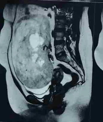
In this case, CT imaging showed a large left ovarian mass, mild ascites, and bilateral pleural effusion, while tumour markers demonstrated markedly elevated LDH with modest CA-125 elevation and normal AFP. During surgery, pelvic fluid and a small peritoneal lesion were also evaluated.
However, the final pathology told a different story. The tumour was a dysgerminoma confined to the left ovary, the capsule remained intact, ovarian surface involvement was absent, peritoneal biopsies and omentum were free of tumour, ascitic/fluid cytology showed no malignant cells, and all 37 regional lymph nodes were negative. The final pathological stage was pT1N0.
This case demonstrates that imaging findings suggestive of Stage III disease do not always represent true metastatic spread, and tumour markers must be interpreted in the context of the specific ovarian tumour type.
- Clinical Insight
Ovarian tumours should be evaluated by a gynecologic oncology team before treatment decisions are made. Imaging, tumour markers, frozen section, and final histopathology together determine the correct stage and need for chemotherapy.
Indian Ovarian Cancer Institute (IOCI), Ahmedabad
Every patient is unique. Treatment recommendations are individualized based on clinical evaluation, imaging, pathology and multidisciplinary discussion.
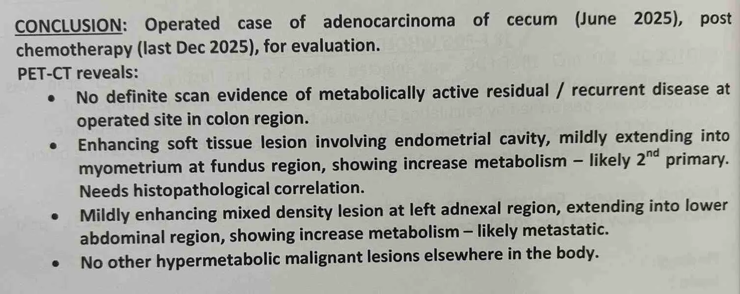
Endometrial Cancer With an Ovarian Mass: Metastasis or a Second Primary?
A 39-year-old woman with a previous history of caecal adenocarcinoma presented with a new endometrial lesion and a large left adnexal mass. She had undergone right hemicolectomy for caecal cancer in June 2025 followed by 8 cycles of CAPOX chemotherapy, completed in December 2025.
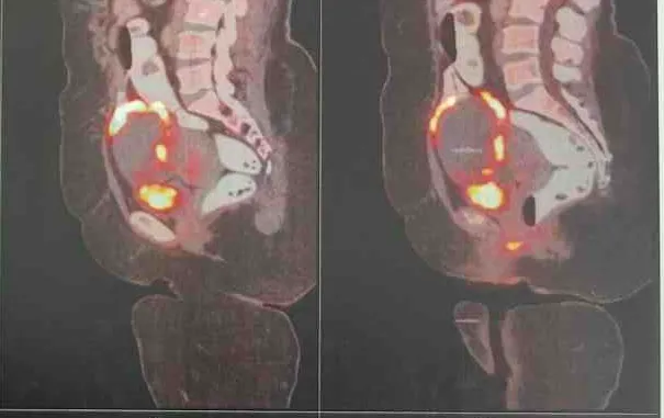
⦿ PET-CT showed an enhancing soft-tissue lesion involving the endometrial cavity, mildly extending into the myometrium at the fundus, with increased radiotracer uptake (SUVmax 17.4). It was reported as likely a second primary and histopathological correlation was advised. A separate mildly enhancing mixed-density lesion measuring approximately 8.5 × 6.9 × 9.0 cm in the left adnexal region, extending into the lower abdomen, demonstrated increased uptake (SUVmax 25.5) and was considered likely metastatic. No other hypermetabolic malignant lesions were identified.
⦿ Endometrial sampling demonstrated endometrioid carcinoma, FIGO Grade II.
⦿ At surgery, a 7 × 7 cm left adnexal mass and bulky right ovary were identified, with no obvious widespread peritoneal disease.
- Clinical Insight
A new ovarian/adnexal mass in a patient with previous cancer cannot automatically be assumed to represent recurrence. Correlation of imaging, pathology, operative findings and molecular information is essential.
Indian Ovarian Cancer Institute (IOCI), Ahmedabad
Every patient is unique. Treatment recommendations are individualized based on clinical evaluation, imaging, pathology and multidisciplinary discussion.
A Large Ovarian Tumour With Endometrial Cancer: Why Complete Staging Matters
In this 39-year-old woman, final histopathology demonstrated endometrioid carcinoma, FIGO Grade 2, involving the endometrium and left ovary.
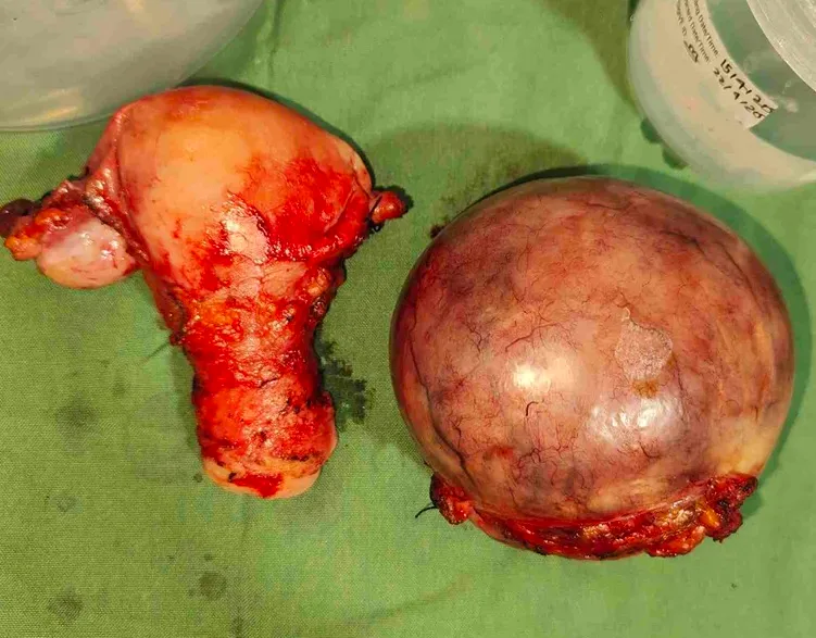
⦿ The separately received left ovarian specimen measured 9.5 × 9 × 8 cm and contained endometrioid carcinoma, with an intact ovarian capsule. Importantly, the omentum and peritoneal biopsies were free of tumour and peritoneal fluid did not contain malignant cells.
⦿ However, systematic lymph-node assessment revealed clinically important occult disease. One of six right pelvic lymph nodes and two of four retroperitoneal lymph nodes contained metastatic macrometastases (>2 mm). Lymphovascular invasion was also present.
⦿ macrometastases (>2 mm). Lymphovascular invasion was also present.
⦿ The final pathological classification was pT3a pN2a, FIGO IIIC2ii.
⦿ Thus, despite the absence of gross peritoneal disease, the patient had significant retroperitoneal nodal involvement. This could only be established through comprehensive surgical staging and pathological examination.
- Clinical Insight
In endometrial cancer with ovarian involvement, a normal-appearing omentum or peritoneum does not exclude advanced disease. Pelvic and retroperitoneal nodal assessment can substantially change the final stage and subsequent treatment strategy.
Indian Ovarian Cancer Institute (IOCI), Ahmedabad
Every patient is unique. Treatment recommendations are individualized based on clinical evaluation, imaging, pathology and multidisciplinary discussion.
Colorectal, Endometrial and Ovarian Cancers in a Young Patient: Why Genetic Testing Was Important
A 39-year-old woman with a previous caecal adenocarcinoma subsequently developed endometrioid carcinoma of the endometrium with ovarian involvement. The occurrence of colorectal and endometrial malignancies at a young age, together with an ovarian endometrioid carcinoma, raised an important possibility of an underlying hereditary cancer syndrome.
Genetic testing identified a heterozygous pathogenic variant in the MSH2 gene: c.1852_1854del (p.Lys618del). The molecular report classified this variant as pathogenic, with autosomal-dominant inheritance, and associated it with Lynch syndrome 2.
This finding provides a unifying explanation for the patient’s pattern of malignancies. Lynch syndrome is particularly relevant when colorectal and endometrial cancers occur at a young age, and ovarian cancer can also occur within the Lynch-associated tumour spectrum.
The importance of identifying an MSH2 pathogenic variant extends beyond the current cancer. It can influence assessment for additional Lynch-associated malignancies, guide tumour molecular evaluation and help determine whether appropriate cascade genetic testing and surveillance should be offered to family members.
In this case, the genetic finding therefore became an important component of the overall oncology assessment rather than being an isolated laboratory result.
- Clinical Insight
A young patient with colorectal cancer followed by endometrial and ovarian malignancy should prompt consideration of hereditary cancer syndromes. Genetic testing can provide information relevant to both the patient’s treatment pathway and the cancer risk assessment of the family.
Indian Ovarian Cancer Institute (IOCI), Ahmedabad
Every patient is unique. Treatment recommendations are individualized based on clinical evaluation, imaging, pathology and multidisciplinary discussion.
Why Immunohistochemistry and Genetic Testing Matter in Endometrial Cancer
⦿ In this 39-year-old woman, histopathology demonstrated endometrioid carcinoma, FIGO Grade 2, involving the endometrium and left ovary, with metastatic macrometastases in pelvic and retroperitoneal lymph nodes. The final pathological stage was pT3a pN2a, FIGO IIIC2ii.
⦿ The pathology report noted that the histomorphology favoured endometrioid carcinoma and that IHC was required for confirmation of the subtype if clinically indicated. This is an important point because modern endometrial cancer assessment increasingly incorporates tumour biology in addition to morphology.
⦿ Immunohistochemistry (IHC) can assess the mismatch-repair proteins MLH1, PMS2, MSH2 and MSH6. Identification of mismatch-repair deficiency (dMMR) has implications for molecular classification, hereditary cancer evaluation and systemic treatment selection.
⦿ In this patient, genetic testing subsequently identified a pathogenic MSH2 variant, c.1852_1854del (p.Lys618del), consistent with Lynch syndrome 2. The patient’s systemic treatment was subsequently planned with chemotherapy combined with dostarlimab, bringing the tumour’s molecular characteristics into the treatment pathway.
⦿ The sequence is clinically important: histopathology → IHC/mismatch-repair assessment → genetic evaluation when indicated → treatment selection.
- Clinical Insight
IHC and molecular testing are not merely additional reports. In appropriately selected endometrial cancer patients, they can identify mismatch-repair abnormalities, uncover hereditary cancer syndromes and help determine whether an immune-checkpoint inhibitor such as dostarlimab should form part of systemic treatment.
Indian Ovarian Cancer Institute (IOCI), Ahmedabad
Every patient is unique. Treatment recommendations are individualized based on clinical evaluation, imaging, pathology and multidisciplinary discussion.
Small Ovarian Cyst, But the Ovary Was Preserved
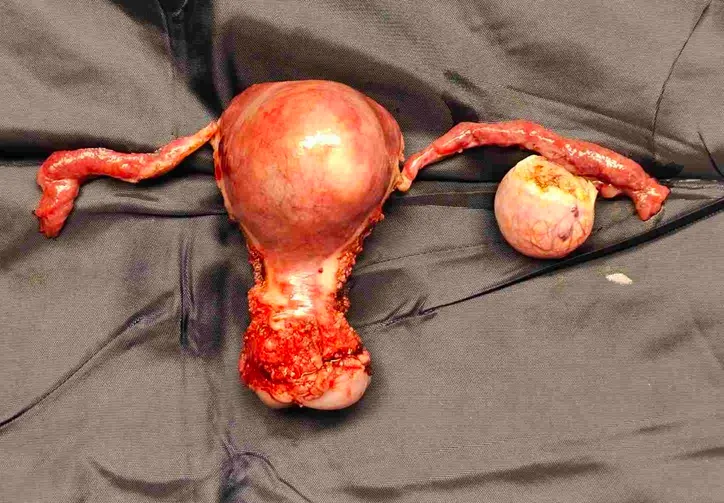
A recently managed 33-year-old woman at IOCI had a small left ovarian/adnexal cyst detected on ultrasound, measuring approximately 2 cm, with internal septations and described as a probable haemorrhagic cyst. The patient also had a posterior subserosal fibroid and a history of atypical endometrial pathology.
During robotic surgery, the left adnexal mass was identified at approximately 3 × 3 cm. Importantly, the mass was resected while preserving the residual healthy ovarian tissue. The right ovary was grossly normal. The uterus and bilateral tubes were removed as planned.
Final histopathology demonstrated a luteal and follicular cyst, with no malignant aetiology.
Key learning:
Finding an ovarian cyst does not automatically mean removing the entire ovary. In appropriately selected young women, ovarian cystectomy with preservation of healthy ovarian tissue can maintain ovarian function while providing definitive tissue diagnosis.
Robotic Ovarian Cyst Removal: Can We Remove the Cyst Intact and Save the Ovary?
⦿ A small ovarian cyst can sometimes create a difficult surgical decision: remove the entire ovary or remove only the lesion?
⦿ In this recently managed IOCI case, robotic surgery allowed precise dissection of the left adnexal mass, separation from surrounding structures and resection of the mass while preserving healthy residual ovarian tissue. The operative findings documented a 3 × 3 cm left adnexal mass, with the opposite ovary grossly normal.
⦿ The specimen was separately submitted for histopathology and measured 2.5 × 2 × 1.5 cm; it was solid-cystic on cut section. Histology subsequently confirmed a luteal and follicular cyst, with no malignancy.
Key learning:
For selected benign-appearing ovarian cysts in young women, the objective should not simply be “remove the ovary.” Intact cyst removal with ovarian preservation can provide diagnosis while retaining functional ovarian tissue.
Robotic Surgery and Early Recovery: Less Surgical Trauma, Faster Mobilisation
⦿ This case demonstrates the practical recovery advantages of a minimally invasive robotic approach.
⦿ The patient underwent hysterectomy with bilateral salpingectomy and left adnexal mass resection along with hysteroscopy/D&C and adhesiolysis. The operation had an estimated blood loss of only 50 mL, with no blood transfusion. The adnexal mass was removed while preserving healthy ovarian tissue.
⦿ Postoperatively, recovery was uneventful. The Foley catheter was removed on postoperative day 1, and the patient was ambulatory, haemodynamically stable, with controlled pain and a clean surgical wound at discharge. She was discharged only a few days after surgery.
Key learning:
Robotic surgery is not only about precision during the operation. In appropriately selected patients, minimally invasive surgery can mean smaller access wounds, low blood loss, early catheter removal, early mobilisation and faster return towards normal activity.
What Are the Real Benefits of Robotic Surgery?
⦿ This recently managed IOCI case demonstrates how robotic technology can facilitate complex pelvic surgery while preserving important structures.
⦿ The procedure involved hysterectomy, bilateral salpingectomy, left adnexal mass resection and adhesiolysis. The surgeon was able to carefully dissect the pelvis, perform bladder dissection, control the uterine vessels and remove the adnexal mass while preserving healthy ovarian tissue. The specimen was delivered vaginally, avoiding an additional abdominal extraction incision.
⦿ The operation was completed with 50 mL blood loss and no transfusion. Postoperative recovery was uncomplicated, with early Foley removal and the patient ambulatory and stable at discharge.
Key learning:
The value of robotic surgery lies beyond the word “robotic.” Magnified 3D vision, wristed instruments, precise tissue dissection and improved pelvic access can help the surgeon perform meticulous minimally invasive surgery—particularly when adhesiolysis, adnexal surgery and pelvic dissection need to be combined.
Get health updates from Dr Swati Shah on WhatsApp
Swasth Parivar: twice a month, practical advice on recovery, diet and warning signs. No advertising. Stop any time.
Your number is used for these updates and for the clinic to reach you. It is never sold or passed on. To stop, reply STOP on WhatsApp.