


Frozen Section
(LIVE Biopsy During Ovarian Tumour Surgery)
What is Frozen Section?
Frozen section is a rapid biopsy done during surgery to diagnose an ovarian cyst, tumour, or mass in real time. In simple terms, it is a “LIVE biopsy” performed while the patient is still in the operation theatre.- Tissue is analysed immediately
- Report is available within 15–30 minutes
- Helps decide the next step of surgery instantly
Dr Swati Shah, MS, DNB — Robotic UroGynec Oncosurgeon, performs frozen-section-guided ovarian tumour surgery at Apollo Hospital, Bhat, Gandhinagar.
What is a frozen section in ovarian surgery?
A frozen section is a rapid microscopic examination of the removed ovarian tissue, done while the patient is still under anaesthesia.
How long does a frozen section take?
The pathologist usually reports within twenty to forty minutes, so the operation continues while the answer is awaited.
How accurate is frozen section for an ovarian tumour?
Frozen section is highly reliable for telling benign from malignant. Borderline tumours are the harder group and need the final histopathology report.
Why does the frozen section matter?
It decides in the same sitting whether removing the mass is enough or whether full staging surgery should be completed straight away.
Who performs frozen-section-guided ovarian surgery in Ahmedabad?
Dr Swati Shah, MS, DNB — Robotic UroGynec Oncosurgeon, performs frozen-section-guided ovarian tumour surgery at Apollo Hospital, Bhat, Gandhinagar.
Does frozen section replace the final biopsy report?
No. Final histopathology follows in three to five days and is the report that confirms the diagnosis and the stage.
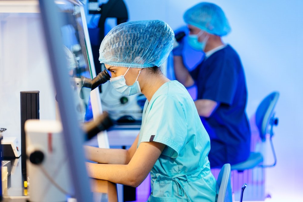
How Frozen Section Works
- Tumour is removed without rupture
- A small sample is sent fresh to the lab
- Tissue is frozen, sliced, and examined under microscope
- Pathologist gives an immediate diagnosis

Why Frozen Section is Critical in Ovarian Tumours
In most ovarian cases:
- Biopsy is NOT done before surgery
- Because it can cause tumour rupture and spillage
That’s where frozen section becomes essential
It acts as a safe “LIVE biopsy” during surgery
 Treatment is not decided only before surgery
 How Treatment is Planned
Final decisions are made during surgery using:
- Resection without rupture
- Frozen section (LIVE biopsy)
This allows:
Right surgery to be performed in a single operation

Why Biopsy Should Be Avoided Before Surgery
In most ovarian masses, preoperative biopsy is NOT recommended
1. Risk of Tumour Rupture
- Needle biopsy can puncture the cyst/tumour
- Leads to spillage → cancer spread
2. Does Not Change Treatment Plan
- If biopsy shows cancer → surgery is still required
- If biopsy shows no cancer → not reliable (only small sample taken)
3. False Reassurance Risk
- A negative biopsy does NOT rule out cancer
Why Not Do Biopsy Before Surgery?
Risk of Biopsy Before Surgery
- Risk of tumour rupture → cancer spread
- May increase cancer stage
- May lead to unnecessary chemotherapy
Why Biopsy is Not Reliable
- A small biopsy sample is not always reliable
- Surgery is still required even if biopsy shows cancer
Therefore, diagnosis is best done during surgery using frozen section
How Frozen Section Changes Surgery (Real Impact)
Frozen section helps answer 3 critical questions immediately:
1. Is it benign / borderline / malignant?
2. What type of tumour is it?
3. How much surgery is needed?
Benign tumour
Limited surgery / ovary preservation
Borderline tumour
Staging with fertility consideration
Malignant tumour
Complete staging / cytoreductive surgery
One surgery. One decision. No delay.
Helps avoid second surgery
When is Frozen Section Used?
Frozen section is commonly used during ovarian tumour surgery to help surgeons make accurate decisions immediately during the operation.
Used in Most Ovarian Masses
Frozen section is used in most localized ovarian cysts, complex cysts, tumours, or masses
Important Principle
Since biopsy is avoided before surgery,
Frozen section becomes the safest and most effective way to diagnose during surgery
Accuracy of Frozen Section
Frozen section provides rapid intraoperative diagnosis and helps surgeons make accurate treatment decisions during ovarian tumour surgery.
Highly Reliable
- Accuracy: ~90–97%
- Highly reliable for benign vs malignant tumours
Limitations
- Borderline tumours
- Very large masses
Final confirmation is always done by histopathology
Frozen Section vs Final Histopathology
Frozen Section
Timing: During surgery
Speed: 15–30 minutes
Purpose: Guide surgery
Detail: Preliminary
Final Histopathology
Timing: After surgery
Speed: Few days
Purpose: Confirm diagnosis
Detail: Detailed
What If Frozen Section is Not Available?
Surgery may be:
Incomplete (miss cancer)
Excessive (remove more than needed)
Or patient may need:
Second surgery
This increases:
Cost
Stress
Treatment delay
Why Frozen Section Requires a Specialized Centre
Needs on-site pathology setup
Requires coordination between
- Surgeon
- Pathologist
Not available at all hospitals
Lets be Informed
Google Reviews
Read real stories from patients across India who trusted our specialized surgical protocols for their recovery.
Mansi Modi
"Dr is very brilliant in her work. Mam operated my mom for ovarian cancer. After two years of operation she guides us so genuinely. Even me also for my regular follow ups. She is so polite. Me and my mom came for follow up regularly and we are very satisfied."
Sharmila Baid
"My search for oncosurgeon ends with Dr swati shah, we came from bikaner Rajasthan for surgery of my sister underwent cytoreductive surgery + Hipec was done by Dr swati shah. She is best gynac cancer surgeon in Ahmedabad Gujarat. We are happy after surgery."
Shradhdhaben Goswami
"My mother underwent laproscopic surgery for ovary cancer and then we found out that it had cancer. The cancer came from appendix and spread to all inside abdomen, omentum and peritoneum cytoreductive surgery and hipec was done by Dr Harsh shah and Dr swati shah. Thank u so much both of you."
Dinesh Patel
"I would like to express my gratitude to Dr swati shah for successful crs + hipec surgery of my mother. We experienced homely care during hospital stay.. Highly recommend Dr swati mam as best ovarian cancer surgeon in Ahmedabad."
jatin trivedi
"CRS+HIPEC surgery was done by Dr swati shah. Her compassionate care and meticulous attention to detail have played crucial role in managing condition. I would like to thanks Dr swati mam for extraordinary effort at all levels. I highly recommend her to anyone in need of exceptional cancer care treatment."
- Knowledge Shorts
Watch our expert-curated shorts for quick, clear answers on diagnosis, treatment & more.
- Video testimonials
Video testimonials feature satisfied patients and clients sharing authentic stories to build trust and credibility.
Ovarian mass resection with frozen section is a specialized surgical procedure performed to remove an ovarian mass, such as a tumor or cyst, from the ovary. During the surgery, a critical step involves sending a small sample of the removed mass for immediate frozen section analysis. This advanced technique involves rapidly freezing the tissue and examining it under a microscope while the surgery is still ongoing. It allows the medical team to immediately determine if the tumor is benign (non-cancerous) or malignant (cancerous), which guides real-time decisions about the extent of the surgery.
During the consultation, it was discovered that the patient had an ovarian cyst that required surgical removal. The laparoscopic procedure was performed just a week before the video was recorded. The patient describes the surgery as a seamless and mostly painless experience. She praises Dr. Swati Shah for her calm, composed nature and exceptional surgical expertise. The minimally invasive nature of the laparoscopy allowed for an incredibly fast recovery; within just one week, the patient’s stitches were removed, and she is already feeling fully recovered and doing well.
- Case Summaries
Exploratory Laparotomy And Complex Excision For A Suspicious Right Adnexal Mass
- The Challenge
- Managing A Suspicious Pelvic Mass
A 49-year-old female presented with severe lower abdominal pain persisting for fifteen days, alongside an alarming heterogenous solid-cystic right pelvic mass. Imaging strongly suggested a neoplastic etiology with pelvic lymphadenopathy, necessitating highly urgent surgical intervention to rule out an aggressive ovarian malignancy.
The Solution
⦿ Comprehensive Exploratory Laparotomy
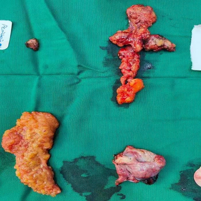
Dr. Swati Shah performed an exploratory laparotomy, right adnexal mass excision, left oophorectomy, appendicectomy, bilateral pelvic lymph node dissection, and omental biopsy. The team navigated complex adhesions, carefully separating the appendix from the tumor. This exceptional surgical precision ensured complete tissue removal and maximized patient safety.
» Surgical Highlights & Precision
• Procedure: An exploratory laparotomy with adnexal excision and appendicectomy was meticulously executed.
• Pelvic Lymph Nodes: Bilateral pelvic lymph node dissection yielded seventeen nodes for comprehensive pathological evaluation.
• Blood Loss: Superior hemostatic control resulted in an exceptionally minimal estimated blood loss of 100 ml.
• Complete Clearance: All suspicious masses and adherent structures were successfully excised without compromising surrounding tissues.
» The Outcome
⦿ Uneventful Recovery And Favorable Pathology
The patient experienced an entirely uneventful postoperative recovery and was discharged with stable hemodynamics. Final histopathology remarkably revealed xanthomatous salpingooophoritis and acute appendicitis, confirming absolutely no malignancy was present. The patient successfully regained ambulatory function and safely transitioned to a comfortable recovery at home.
» Dr. Swati Shah’s Insight
Navigating a highly suspicious pelvic mass with severe adhesions requires absolute surgical precision. By comprehensively excising the involved tissues, we achieved a definitive benign diagnosis, securing the patient's excellent long-term health.
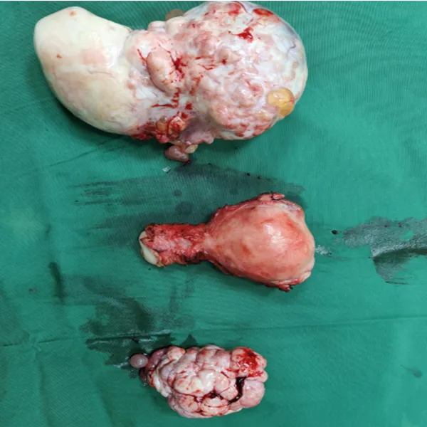
Advanced Surgical Management of Metastatic Ovarian Carcinoma Secondary to Breast Cancer
- The Challenge
- Complex Metastatic Presentation
A 45-year-old female presented with bilateral adnexal masses, signaling a metastatic spread from a previously treated left breast carcinoma. The case was particularly complex due to the patient’s extensive history of prior chemotherapy, mastectomy, and the aggressive, recurrent nature of the metastatic disease requiring urgent intervention.
The Solution
⦿ Comprehensive Exploratory Laparotomy and Multimodal Cytoreduction
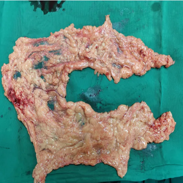
Dr. Swati Shah performed a meticulous diagnostic laparoscopy followed by a formal exploratory laparotomy, total abdominal hysterectomy, and bilateral salpingo-oophorectomy. Precision in surgical technique was paramount to safely resect the adnexal masses while performing an infracolic omentectomy, ensuring the complete removal of all visible metastatic deposits within the abdominal cavity.
» Surgical Highlights & Precision
• Procedure: A comprehensive cytoreductive surgery involving diagnostic laparoscopy, laparotomy, hysterectomy, and infracolic omentectomy was successfully performed.
• Anatomical Complexity: The surgical team navigated challenging bilateral adnexal masses to ensure complete tumor excision from the ovaries.
• Safety & Hemostasis: Exceptional surgical precision was maintained throughout the procedure, resulting in minimal blood loss of 100 ml.
• Cancer Clearance: The procedure achieved a successful oncological outcome with final pathology confirming the removal of all involved tissues.
» The Outcome
⦿ Stable Recovery and Pathological Success
Postoperatively, the patient remained hemodynamically stable and was mobilized shortly after the intervention. Pathological evaluation confirmed the successful resection of metastatic carcinoma with clear margins, while the uterus and parametria remained uninvolved. The patient experienced an uneventful recovery and was discharged in stable condition to continue her ongoing oncological management.
» Dr. Swati Shah’s Insight
In cases of metastatic breast cancer involving the ovaries, a meticulous surgical approach is vital to managing the disease burden effectively. By utilizing a comprehensive cytoreductive strategy, we can achieve optimal outcomes and provide our patients with the best chance for long-term survival.
Consult for Expert Ovarian Tumour Surgery
Proper planning with frozen section guidance helps achieve safer and more accurate treatment decisions.
If you have:
- Ovarian cyst
- Complex ovarian mass
- Suspicion of ovarian cancer
Frozen section availability can help ensure:
- Accurate diagnosis
- Right extent of surgery
- Better outcomes
Frequently Asked Questions
A rapid test done during surgery to diagnose the tumour immediately.
Yes, it is highly accurate (~90–97%), but final confirmation is done later.
No. It takes about 15–30 minutes and helps guide proper surgery.
It is used in most suspicious or unclear ovarian tumours.
Yes, it helps identify cancer during surgery and guides treatment.
Get health updates from Dr Swati Shah on WhatsApp
Swasth Parivar: twice a month, practical advice on recovery, diet and warning signs. No advertising. Stop any time.
Your number is used for these updates and for the clinic to reach you. It is never sold or passed on. To stop, reply STOP on WhatsApp.